T4 [FBbt_00003731]


T4
ID: FBbt_00003731
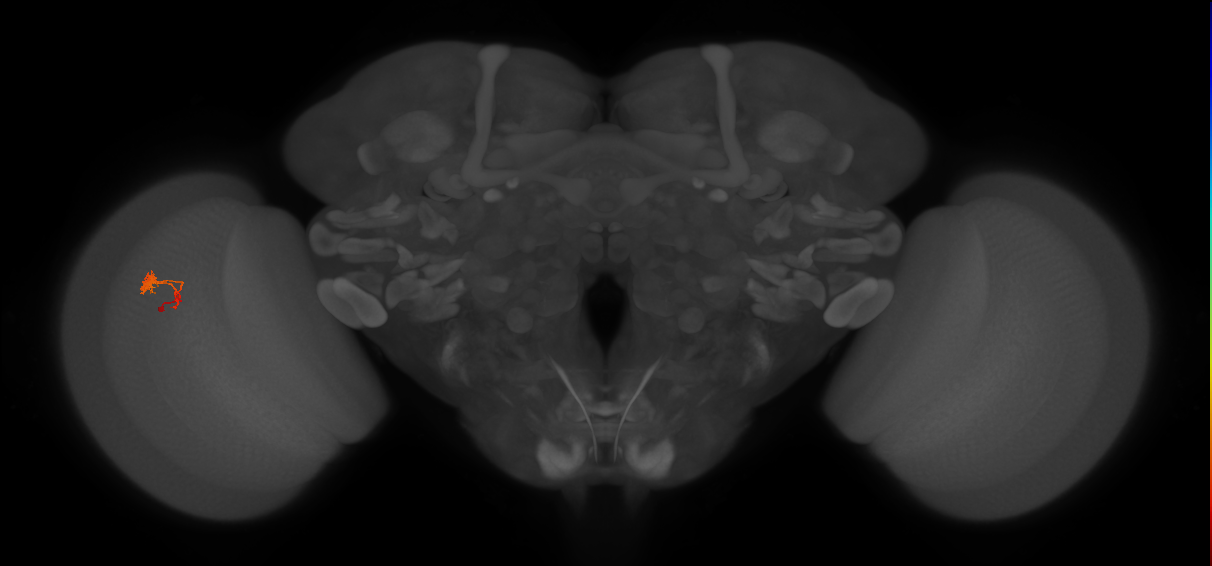
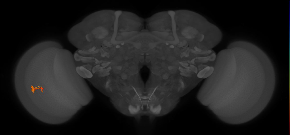
T neuron with its cell body posterior to the lobula plate and a cell body fiber that projects through the lobula plate and via the second optic chiasma, with little or no arborization, to medulla layer 10 (Fischbach and Dittrich, 1989; Shinomiya et al., 2019). It forms a fine arborization in medulla layers M9 and M10 and its axon then doubles back across the second optic chiasma to form bleb-type arborizations in a single layer of the lobula plate (Fischbach and Dittrich, 1989; Shinomiya et al., 2019). It is a cholinergic neuron (Mauss et al., 2014; Shinomiya et al., 2014). There are a large number of these cells, which are generated from neuroblasts that amplify by symmetric division (type III) before generating the T4 (and T5) neurons (Mora et al., 2018).
Pre- versus postsynaptic innervation judged by scoring of terminal morphology from figures in Fischbach and Dittrich (1989) as assessed by FlyBrain Neuron DB. The neurotransmitter was assessed with a split-GAL4 strategy using a Chat-DBD allele (FBal0198074) (Mauss et al., 2014) and single-cell transcript profiling (Shinomiya et al., 2014).
Open in VFB 3D Browser →Relationships
- develops from: immature T4 neuron
- has soma location: cell body rind of adult posterior lobula plate
- receives synaptic input in region: medulla layer M10, medulla layer M9
- sends synaptic output to region: lobula plate layer
Alternative Names
| Synonym | Scope | Reference |
|---|---|---|
| T4 | exact synonym | Özel et al., 2021 |
| T neuron T4 | exact synonym |
Feedback
Was this page helpful?
Glad to hear it! Please tell us how we can improve.
Sorry to hear that. Please tell us how we can improve.