medulla sublayer M3A [FBbt_00111267]
Distal sublayer of the medulla layer M3, proximal to layer M2. It is defined by the extent of the arborization of Dm3 neurons.
medulla sublayer M3A
ID: FBbt_00111267
Adult Nervous system Synaptic neuropil subdomain Visual system
Distal sublayer of the medulla layer M3, proximal to layer M2. It is defined by the extent of the arborization of Dm3 neurons.
In the literature there are two methods for describing spatial location of neurons in the visual system: distal-proximal (e.g. Fischbach and Dittrich, 1989), and medial-lateral (e.g. Ito et al., 2014), both meaning outer and inner, and refer to the distance of a structure along the visual pathway relative to the center of the brain. Currently we will adopt the classical 'distal-proximal'.
Open in VFB 3D Browser →Relationships
- is part of: medulla layer M3
Neurons with some part in medulla sublayer M3A (4 total)
| Thumbnail | Name | Tags |
|---|---|---|
 |
Dm3 | Adult, Glutamatergic, Nervous_system, Visual_system, secondary_neuron |
 |
Dm3a | Adult, Glutamatergic, Nervous_system, Visual_system, secondary_neuron |
 |
Dm3b | Adult, Glutamatergic, Nervous_system, Visual_system, secondary_neuron |
 |
Dm3c | Adult, Glutamatergic, Nervous_system, Visual_system, secondary_neuron |
Neurons with synaptic terminals in medulla sublayer M3A (4 total)
| Thumbnail | Name | Tags |
|---|---|---|
|
Dm3 | Adult, Glutamatergic, Nervous_system, Visual_system, secondary_neuron |
|
Dm3a | Adult, Glutamatergic, Nervous_system, Visual_system, secondary_neuron |
|
Dm3b | Adult, Glutamatergic, Nervous_system, Visual_system, secondary_neuron |
|
Dm3c | Adult, Glutamatergic, Nervous_system, Visual_system, secondary_neuron |
Neurons with presynaptic terminals in medulla sublayer M3A (4 total)
| Thumbnail | Name | Tags |
|---|---|---|
|
Dm3 | Adult, Glutamatergic, Nervous_system, Visual_system, secondary_neuron |
|
Dm3a | Adult, Glutamatergic, Nervous_system, Visual_system, secondary_neuron |
|
Dm3b | Adult, Glutamatergic, Nervous_system, Visual_system, secondary_neuron |
|
Dm3c | Adult, Glutamatergic, Nervous_system, Visual_system, secondary_neuron |
Neurons with postsynaptic terminals in medulla sublayer M3A (4 total)
| Thumbnail | Name | Tags |
|---|---|---|
|
Dm3 | Adult, Glutamatergic, Nervous_system, Visual_system, secondary_neuron |
|
Dm3a | Adult, Glutamatergic, Nervous_system, Visual_system, secondary_neuron |
|
Dm3b | Adult, Glutamatergic, Nervous_system, Visual_system, secondary_neuron |
|
Dm3c | Adult, Glutamatergic, Nervous_system, Visual_system, secondary_neuron |
Images of neurons with some part in medulla sublayer M3A (8548 total)
| Thumbnail | Name | Tags |
|---|---|---|
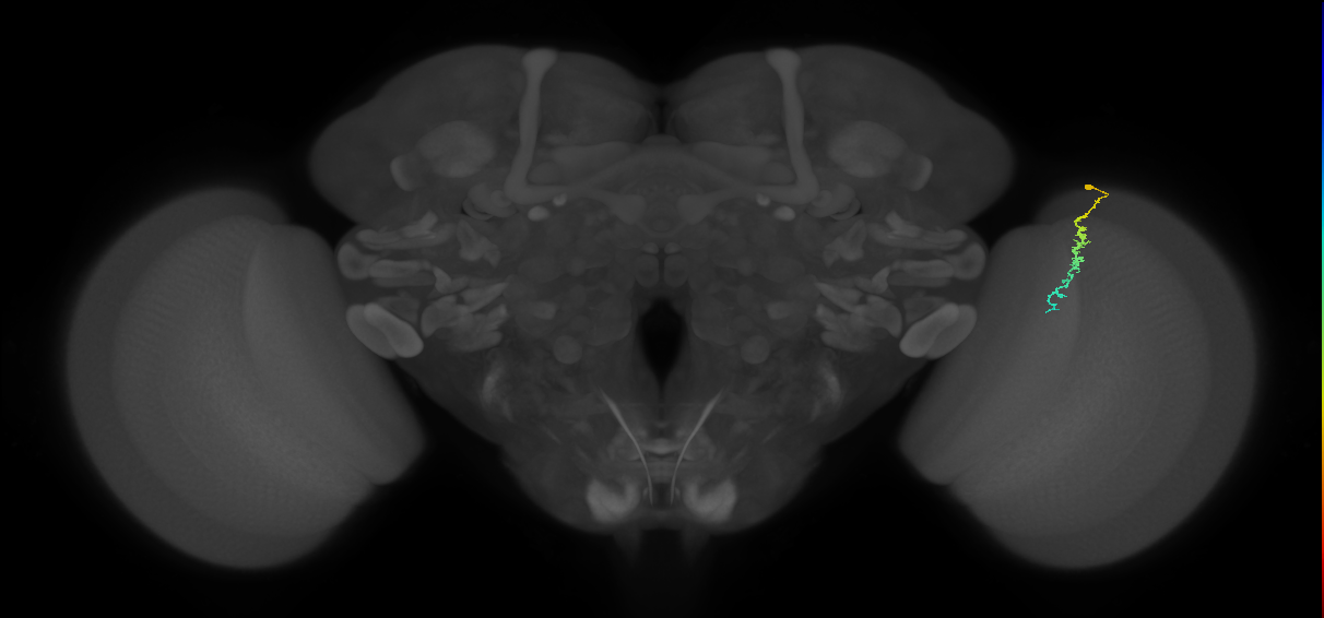
| Dm3a_R (MaleCNS:126069) | Adult, Glutamatergic, Nervous_system, Visual_system, secondary_neuron | |
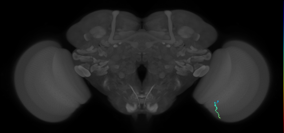
| Dm3a_R (MaleCNS:128302) | Adult, Glutamatergic, Nervous_system, Visual_system, secondary_neuron | |
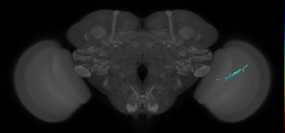
| Dm3c_L (MaleCNS:178458) | Adult, Glutamatergic, Nervous_system, Visual_system, secondary_neuron | |
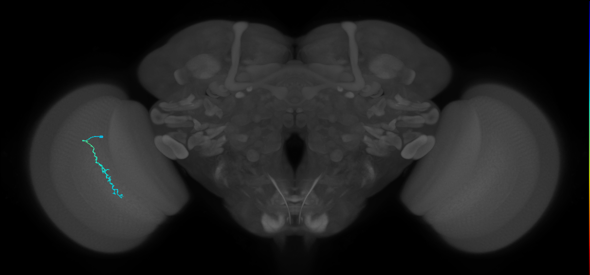
| Dm3c_L (MaleCNS:535894) | Adult, Glutamatergic, Nervous_system, Visual_system, secondary_neuron | |
| Dm3c_R (MaleCNS:153530) | Adult, Glutamatergic, Nervous_system, Visual_system, secondary_neuron |
View all 8548 results in VFB →
Feedback
Was this page helpful?
Glad to hear it! Please tell us how we can improve.
Sorry to hear that. Please tell us how we can improve.