photoreceptor cell of Bolwig organ [FBbt_00007253]

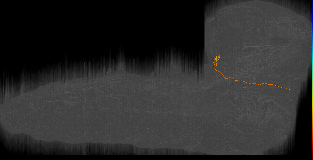
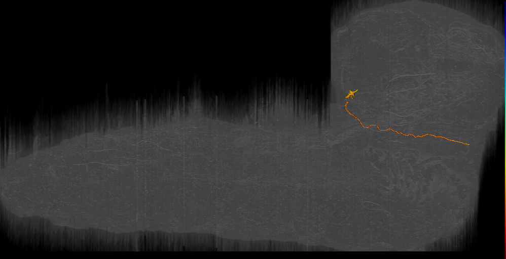
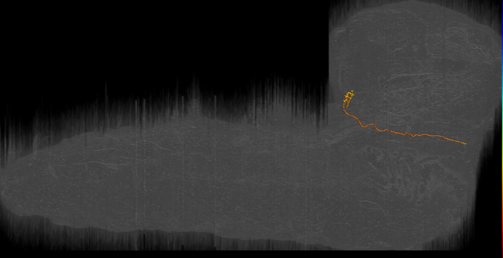
photoreceptor cell of Bolwig organ
ID: FBbt_00007253
Photoreceptor cell that is part of a Bolwig's organ. Unlike the photoreceptors of the adult eye, eyelet and ocellus, these photoreceptors do not contain rhabdomeres, but instead have apical surfaces which are folded into numerous horizontal microtubule-containing microvillar-like processes (Green et al., 1993; Hartenstein et al., 2019). These processes are variable in length, diameter and spacing, but align approximately along a ventro-anterior to dorso-posterior axis relative to the body and do not branch (Hartenstein et al., 2019). There are around 12 of these cells per hemisphere and their axons enter the brain ventrolaterally via the Bolwig nerve and terminate in the larval optic neuropil (Larderet et al., 2017). There are two mutually exclusive subtypes, expressing Rh5 or Rh6, both of which are cholinergic and are capable of entraining the molecular clock by light (Keene et al., 2011).
Open in VFB 3D Browser →- embryonic/larval sensory neuron
- larval PNS neuron
- larval cholinergic neuron
- photoreceptor neuron
- sensory organ cell
Relationships
- bearer of: unilateral
- capable of: entrainment of circadian clock by photoperiod
- fasciculates with: Bolwig nerve
- has soma location: Bolwig organ
- sends synaptic output to region: larval optic neuropil
Alternative Names
| Synonym | Scope | Reference |
|---|---|---|
| larval photoreceptor neuron | exact synonym | Larderet et al., 2017 |
| larval PRC | exact synonym | Hartenstein et al., 2019 |
| photoreceptor cell of Bolwig’s organ | exact synonym |
Feedback
Was this page helpful?
Glad to hear it! Please tell us how we can improve.
Sorry to hear that. Please tell us how we can improve.