VFB hosts a range of datatypes, including electron microscopy (EM) and light microscopy (LM) datasets with images annotated using the same cell type ontology terms and aligned to common standard templates. We also host single cell RNA sequencing (scRNAseq) gene expression data.
This is the multi-page printable view of this section. Click here to print.
Data on VFB
Types of data and datasets that are available on Virtual Fly Brain.
- 1: Electron Microscopy Data
- 2: Light Microscopy Data
- 2.1: Janelia FlyLight
- 3: Single Cell RNA Sequencing Data
- 4: Templates
1 - Electron Microscopy Data
Electron Microscopy Data available on Virtual Fly Brain.
Virtual Fly Brain brings together data from multiple electron microscopy (EM) resources, providing access to high-resolution neuroanatomical datasets. These datasets include complete EM volumes of Drosophila brains and ventral nerve cords, neuron reconstructions, connectivity information and neurotransmitter predictions. Data can be visualised using the web browser or accessed programmatically via APIs.
Comparison Table of Integrated Datasets
The table below summarises EM datasets that have been integrated into VFB, including the portion of the organism covered (Anatomy), the resource(s) where the original data can be found, The level of reconstruction (sparse or dense) and the original publication for the dataset.
| Dataset | VFB symbol | Anatomy | Reconstruction | Resource(s) | Original Publication |
|---|---|---|---|---|---|
| BANC | BANC | Full CNS (adult female) | Dense | Codex | Bates et al. (2025) |
| male-CNS | mc | Full CNS (adult male) | Dense | NeuPrint; Codex | Berg et al. (2025) |
| Optic-lobe | ol | Optic lobe (adult male) | Dense | NeuPrint; Codex | Nern et al. (2025) |
| FAFB (FlyWire) | fw | Full brain (adult female) | Dense | Codex | Dorkenwald et al. (2024); Schlegel et al. (2024) |
| MANC | mv | Full VNC (adult male) | Dense | NeuPrint; Codex | Takemura et al. (2024) |
| FANC | N/A | Full VNS (adult female) | Sparse | CATMAID | Phelps et al. (2021) |
| Hemibrain | hb | Partial brain (adult female) | Dense | NeuPrint | Scheffer et al. (2020) |
| FAFB (CATMAID) | fafb | Full brain (adult female) | Sparse | CATMAID | Zheng et al. (2018) |
| l1em | l1em | Full CNS (female larva) | Sparse | CATMAID | Ohyama et al. (2015) |
Datasets Hosted by VFB
Virtual Fly Brain (VFB) hosts several CATMAID instances for exploring connectomic reconstruction data. These datasets provide access to neuroanatomical data from various Drosophila electron microscopy projects. Full details are available here.
1.1 - Dataset Versions and Deprecation
How Virtual Fly Brain handles neurons, connectivity, data sources and cross-references when new versions of connectomic datasets are released.
Connectomic datasets are not static: reconstructions are re-released as proofreading improves, neurons are split, merged or re-identified, and the resources that host them may occasionally move or shut down. When VFB ingests a new release, it must decide what happens to the entities from the previous version. This page documents those policies so that users understand why a neuron, link or connection may change, disappear, or persist between releases.
Affected entities
A connectomic dataset is represented in VFB by several types of graph node and edge:
| Node / edge | What it represents |
|---|---|
| DataSet nodes | A specific released version of a dataset (e.g. a FlyWire release). Neurons are attached to it via has_source. |
| Site (data source) nodes | The external resource that hosts the data (e.g. Codex, NeuPrint, CATMAID). Holds the link_base used to build cross-reference links, and is flagged is_data_source = [true] when it is the canonical source for its neurons. |
| Neuron (Individual) nodes | A single reconstructed neuron, cell typed by linking to anatomy ontology (FBbt) nodes via INSTANCEOF edges, with other annotations (soma location, developmental origin, sex, etc.) linked via other edge types. |
| Image / Channel nodes | The Neuron’s aligned image(s) and the channel(s) registered to a template. |
| Connectivity edges | Synaptic connectivity (synapsed_to) between Neurons. |
Cross-reference edges (database_cross_reference) |
Links a Neuron to a Site, carrying the accession (the Neuron’s ID in that resource). |
Core principle: Site and Neuron deprecation are independent
Whether a Site is deprecated and whether a Neuron is deprecated are decided separately. A Neuron can be valid while its data source is deprecated, and a data source can remain live while individual Neurons within it are retired. The two states are tracked independently and have different consequences (below).
Deprecation does not delete a node. The node and its identifier are retained (so old IDs resolve), but it is marked deprecated and treated accordingly.
What happens when a new dataset version is released
For example, when BANC v626 (the old version) is replaced by v888 (the new version):
- New nodes are created for the new Site and DataSet, and for any new Neurons in the release (accessions that were not previously in VFB). The markers that identify a Site as the live, canonical connectome source — its symbol (the short dataset code listed in EM Data),
Connectomelabel andis_data_sourceflag — are transferred from the old Site to the new one (removed from the old, added to the new), so the new Site takes over as the canonical source. - A
term_replaced_byedge is added to link the old and new DataSet/Site. - Old DataSet — deprecated (superseded by the new DataSet, which it links to via
term_replaced_by). - Old Sites that still exist, i.e. links will still resolve — not deprecated.
- Old Sites that no longer exist, i.e. links will not resolve — deprecated.
- Neurons with accessions that persist in the new data — not deprecated. They keep their
database_cross_referenceedge to the old Site and gain an edge to the new Site. - Neurons with accessions that are not present in the new data — deprecated. They have no
database_cross_referenceedge to the new Site; their only cross-reference is to the old, deprecated Site. - Connectivity edges are replaced with edges from the new data for Neurons that are not deprecated.
- Old images are removed. Images from the new data are loaded for Neurons that are not deprecated.
- Cell type / FBbt links are replaced with annotations based on the new data for Neurons that are not deprecated. These may be removed from deprecated Neurons if they are incorrect based on the new data, or retained if they are still valid.
- Channel nodes are always retained (not deprecated). Deprecation is not necessary, as all queries are keyed off of the Neuron node.
What happens when a dataset/site is retired with no replacement
The Site is deprecated and there is no new Site.
- Neurons — remain valid (not deprecated) and remain valid query targets. Their only data source is now a deprecated Site, so no link can be built to a live resource, but the Neurons, their connectivity and their images are still served.
- The Site — deprecated, but its
is_data_sourceflag remains[true]so the Site stays the canonical source for its Neurons. That attribution is what keeps those Neurons discoverable: they continue to be picked up by queries even though their Site is deprecated. - The DataSet — not deprecated: with no replacement version to supersede it, it remains the current representation of the data.
Effects on the website and queries
These follow from the states above and are enforced when results are generated:
- Deprecated Neurons are excluded from connectivity results and from the Neuron counts used in connectivity summaries (so they do not appear as partners and do not affect percentages).
- Deprecated Sites never produce a clickable external link. Where a results table has source / accession columns (e.g. instance and similar-neuron tables, connectivity tables), the source name and accession are still shown as plain text — they are just not linked. In the Term Info cross-reference list, whose entries exist specifically to be links, a deprecated Site’s entry is omitted.
- Neurons whose only data source is a deprecated Site remain valid query targets and are returned normally; only their outbound link to the dead resource is suppressed.
Technical note
Both is_data_source and the deprecation flag are stored as list-valued annotations in the
database (e.g. is_data_source = [true]). Deprecation also surfaces in the search index as
Deprecated within the node’s types list. Result-generating code keys off these
annotations to apply the behaviours above; Site and Neuron deprecation are checked
independently.
See also
2 - Light Microscopy Data
Light Microscopy Data available on Virtual Fly Brain.
Virtual Fly Brain brings together data from multiple Light Microscopy (LM) resources, providing access to visualisations of driver (including split) expression patterns.
2.1 - Janelia FlyLight
The FlyLight Project produces large anatomical data sets and highly characterized collections of GAL4, LexA and Split-GAL4 drivers in order to enable the visualization and precise manipulation of individual cell types in the Drosophila nervous system.
Introduction
FlyLight makes its primary data available via VFB as well as through dedicated websites for expression patterns of Generation 1 GAL4 and LexA lines and for split-GAL4 lines. Generation 1, split hemidriver, and stable split combination lines are available primarily from the Bloomington Stock Center. Stable split lines can also be requested via the split-GAL4 website.
FlyLight Data and Resources
FlyLight data is integrated and indexed on VFB and direct linkouts are provided from the Term Info tabs to the original images. VFB hosts the original larval images here. External links to the original collections of adult images are provided below:
Generation 1 GAL4 expression patterns
Generation 1 MCFO (single neuron) patterns
Split-GAL4 Expression Patterns
Light and EM neurons can also be searched for on NeuronBridge
Also see FlyLight Imaging Tile Documentation.
3 - Single Cell RNA Sequencing Data
scRNAseq Data available on Virtual Fly Brain.
Virtual Fly Brain incorporates scRNAseq data from multiple studies, including the Fly Cell Atlas, Aging Fly Cell Atlas and other datasets that identify nervous system cell types - full list here. These datasets can be found by searching for ‘scRNAseq’ and filtering to ‘Dataset’.
Clusters can be found for particular cell types via the Single cell transcriptomics data for.. query on the cell type Term Info pane.

Genes for a particular cluster can be filtered by function and sorted by expression level and extent (proportion of cells in cluster expressing the gene). As with other VFB search results, these can be exported as a csv.
Note that we currently only include genes that have extent > 0.2 in a cluster.
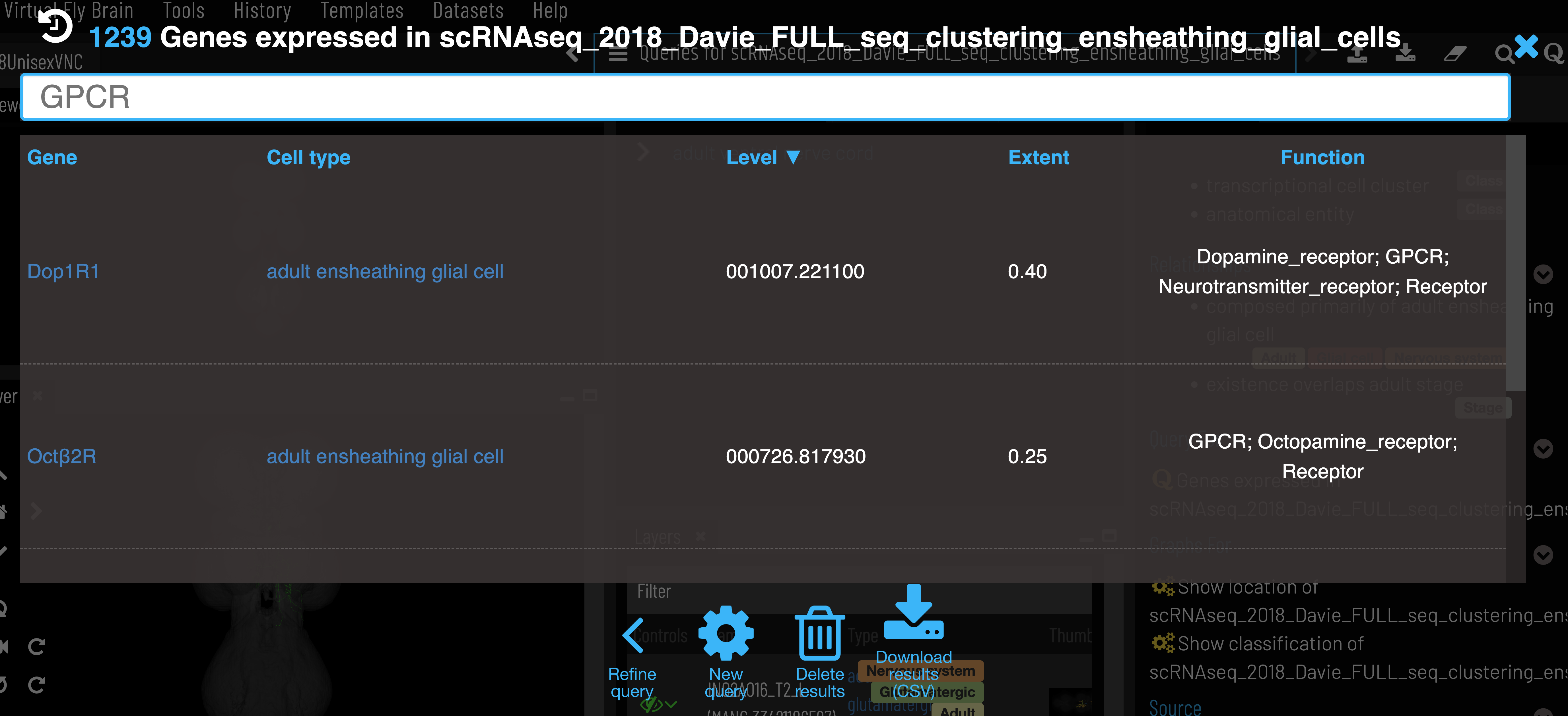
Data can also be retrieved using VFB_connect.
We pull scRNAseq data from FlyBase, which takes it from the Single Cell Expression Atlas.
4 - Templates
Summary of templates on Virtual Fly Brain and the painted domains/regions available in those templates.
Canonical templates not only allow for spatial alignment of image data, but also are often painted to make a reference atlas of anatomical regions. Many central nervous system (CNS) templates exist for Drosophila, below we provide a summary of those used in VFB. Templates can be downloaded from their Term Info pages.
Central Nervous System from Janelia Research Campus JRC2018
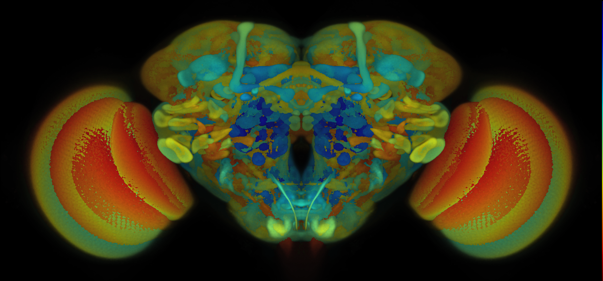
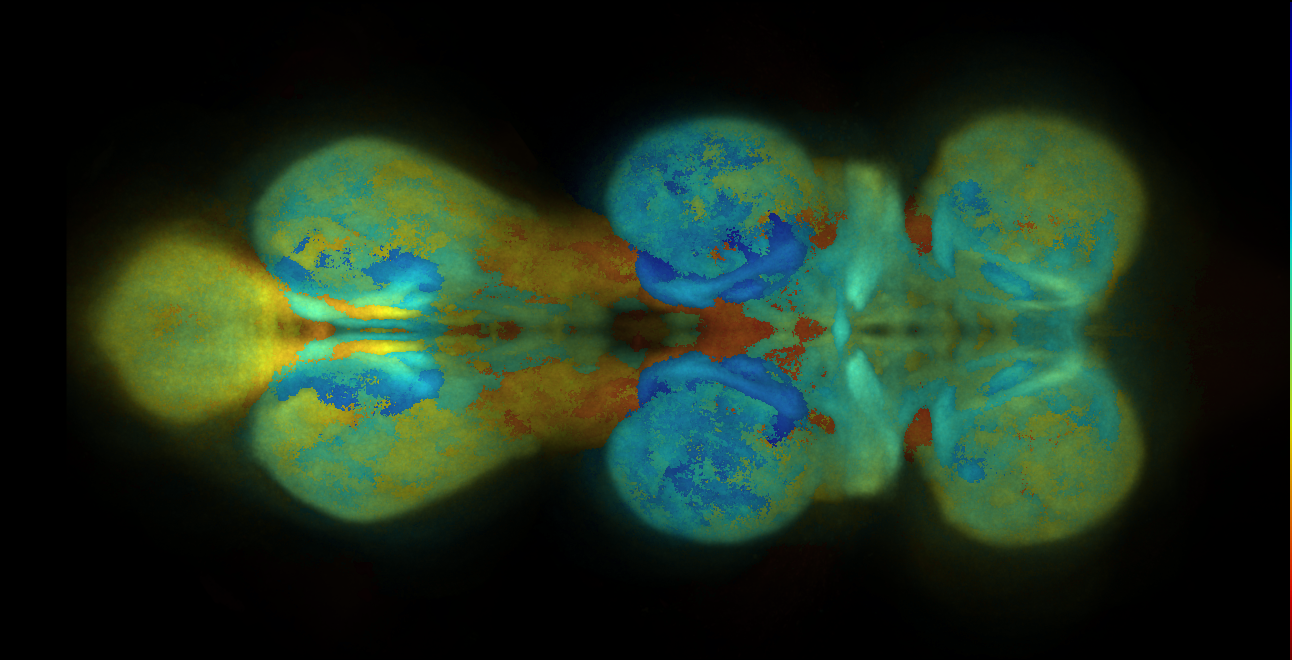
VFB displays data aligned to the JRC 2018 unisex brain template or JRC 2018 unisex Ventral Nerve Cord (VNC), as appropriate. See Bogovic et al., “An unbiased template of the Drosophila brain and ventral nerve cord” for details of template generation.
The original files are available from Janelia: JRC 2018 Brain templates.
Adult Brain from Janelia Research Campus/VFB (JFRC2010/JFRC2)
Superceeded by JRC2018
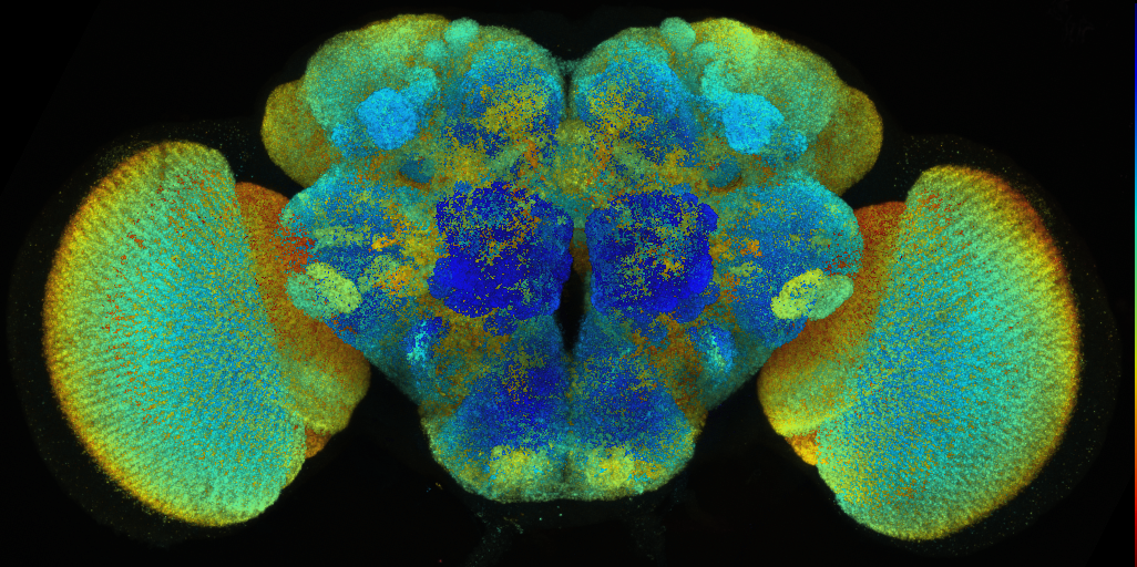
Template brain created by Arnim Jenett (Janelia Research Campus), Kazunori Shinomiya and Kei Ito (Tokyo University) from a staining with the neuropil marker nc82.
The voxel size is 0.62x0.62x0.62 microns.
The files are available here.