DN1a FBbt_00007429

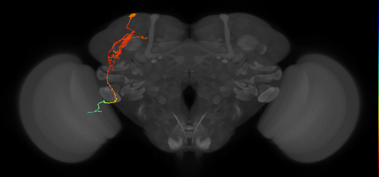
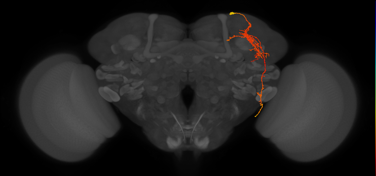

DN1a
ID: FBbt_00007429
Adult DN1 neuron with its soma in the anterior superior brain, dorsal to the superior lateral protocerebrum (SLP) (Shafer et al., 2006; Reinhard et al., 2022. It is glutamatergic and also expresses Nplp1 (IPNamide) and CCHamide1 (Shafer et al., 2006; Collins et al., 2012; Fujiwara et al., 2018; Reinhard et al., 2022 and Dh44 (Reinhard et al., 2024. It arborizes ipsilaterally in the ventromedial posterior lateral horn and the lateral and dorsal mushroom body accessory calyces with mixed pre- and post-synaptic terminals (Bates et al., 2020; Marin et al., 2020; Reinhard et al., 2022. A ventral projection extends to the posterior optic commissure and follows it laterally, reaching the accessory medulla, where it has mainly presynaptic terminals (Bates et al., 2020; Marin et al., 2020; Reinhard et al., 2022. A large amount of input is from the thermosensory VP3 vPN and VP2 adPN neurons (Marin et al., 2020; Alpert et al., 2020. Activity of the cold-responsive VP3 vPN has a strong inhibitory effect on DN1a (Alpert et al., 2020. There are two of these cells per hemisphere and they are primary neurons (Shafer et al., 2006; Bates et al., 2020. One of these cells per hemisphere additionally extends a branch dorsally into the SLP (Reinhard et al., 2022. (Fujiwara et al., 2018, Marin et al., 2020, Collins et al., 2012, Reinhard et al., 2024, Bates et al., 2020, Reinhard et al., 2022, Alpert et al., 2020, Shafer et al., 2006, Baggerman et al., 2002)
Open in VFB 3D Browser →Classification
- CCHamide-1 neuron
- DN1 neuron
- accessory medulla neuron
- adult Dh44 neuron of brain
- adult glutamatergic neuron
- adult lateral horn neuron
- primary neuron
Relationships
- capable_of_part_of: thermosensory behavior (PMID:21195773, PMID:32442464)
- capable_of_part_of: circadian rhythm (PMID:21195773, PMID:32442464)
- bearer of: unilateral
- expresses: Nplp1
- fasciculates with: posterior optic commissure
- has soma location: cell body rind of adult dorsal superior lateral protocerebrum
- receives synaptic input from neuron: adult antennal lobe projection neuron VP2 adPN, adult antennal lobe projection neuron VP3 vPN
- receives synaptic input in region: adult mushroom body dorsal accessory calyx, adult mushroom body lateral accessory calyx
- sends synaptic output to region: accessory medulla, adult lateral horn, adult mushroom body dorsal accessory calyx, adult mushroom body lateral accessory calyx
Alternative Names
| Synonym | Scope | Reference |
|---|---|---|
| DN1a | related synonym | Berg et al., 2025, Bates et al., 2025, name_in_banc, Nern et al., 2025, Schlegel et al., 2024, Scheffer et al., 2020 |
| anterior DN1 neuron | exact synonym | Reinhard et al., 2024 |
| DN1a | exact synonym | Bates et al., 2020 |
Downloads
Image files aligned to DN1a_R:
Image files aligned to DN1a_L (MaleCNS:516397):
Image files aligned to DN1a_R (MaleCNS:21316):
Image files aligned to DN1a_R:
Image files aligned to DN1a:
Image files aligned to DN1a:
Image files aligned to DN1a:
Image files aligned to DN1a_R (JRC_OpticLobe:21316):
Image files aligned to DN1a#2_L (FAFB:9362353):
Image files aligned to DN1a#1_L (FAFB:8867611):