This is the multi-page printable view of this section. Click here to print.
Datasets available on VFB
- 1: Adult Brain fru clones (Cachero2010) [Cachero2010]
- 2: Adult VNC neuropils (Court2020) [Court2020]
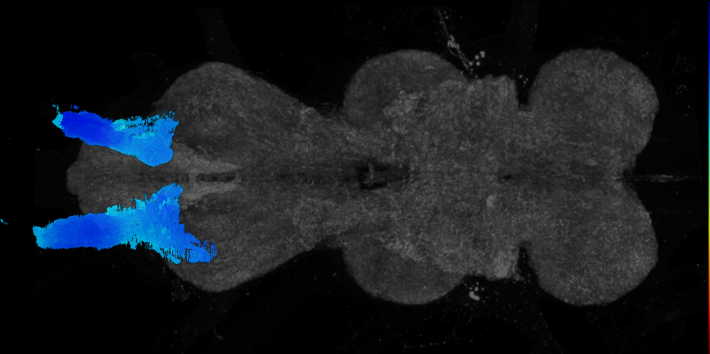
- 3: Adult VNS neuropils (Court2017) [Court2017]
- 4: AMMC local and projection neurons (Matsuo2016) [Matsuo2016]
- 5: banc [Bates2025]
- 6: Biomechanical origins of proprioceptive maps in the Drosophila leg [Mamiya2022]
- 7: BrainName neuropils and tracts - Ito half-brain [BrainName_Ito_half_brain]
- 8: BrainName neuropils on adult brain JFRC2 (Jenett, Shinomya) [JenettShinomya_BrainName]
- 9: BrainTrap lines (Knowles-Barley2010) [Knowles_Barley2010]
- 10: central brain neurons by lineage, Lee2020 [Lee_Lineage2020]
- 11: Comparative Connectomics Reveals How Partner Identity, Location, and Activity Specify Synaptic Connectivity in Drosophila (Valdes-Aleman et al. 2021) [Valdes_Aleman2021]
- 12: Dickson lab VT line collection - VDRC images [Dickson_VT]
- 13: Dickson lab VT lines - FlyLight/Janelia images (2017) [Dickson2017]
- 14: EM FAFB Baltruschat et al 2021 [Baltruschat2021]
- 15: EM FAFB Bates and Schlegel et al 2020 [BatesSchlegel2020]
- 16: EM FAFB Coates et al 2020 [Coates2020]
- 17: EM FAFB Dolan and Belliart-Guerin et al. 2018 [Dolan2018]
- 18: EM FAFB Dolan et al. 2019 [FafbDolan2019]
- 19: EM FAFB Dombrovski et al 2023 [Dombrovski2023]
- 20: EM FAFB Engert et al. 2022 [Engert2022]
- 21: EM FAFB Erginkaya et al 2025 [Erginkaya2025]
- 22: EM FAFB Felsenberg et al. 2018 [Felsenberg2018]
- 23: EM FAFB Gorko et al 2024 [Gorko2024]
- 24: EM FAFB Hampel and Eichler et al 2020 [HampelEichler2020]
- 25: EM FAFB Kim et al 2020 [Kim2020]
- 26: EM FAFB Kind et al. 2021 [Kind2021]
- 27: EM FAFB Marin et al 2020 [Marin2020]
- 28: EM FAFB Morimoto et al 2020 [Morimoto2020]
- 29: EM FAFB Otto et al 2020 [Otto2020]
- 30: EM FAFB Sayin et al 2019 [Sayin2019]
- 31: EM FAFB Shiu et al. 2022 [Shiu2022]
- 32: EM FAFB Taisz and Galili et al., 2022 [TaiszGalili2022]
- 33: EM FAFB Turner-Evans et al 2020 [Turner_Evans2020]
- 34: EM FAFB Wang et al 2020a [Wang2020a]
- 35: EM FAFB Wang et al 2020b [Wang2020b]
- 36: EM FAFB Wang et al 2020c [Wang2020c]
- 37: EM FAFB Zhao et al., 2023 [Zhao2023]
- 38: EM FAFB Zheng et al 2018 [Zheng2018]
- 39: EM FAFB Zheng et al 2020 (published 2022) [Zheng2020]
- 40: EM FANC Phelps et al 2020 [Maniates_Selvin2020]
- 41: EM L1 Andrade et al. 2019 [Andrade2019]
- 42: EM L1 Barnes et al., 2022 [Barnes2022]
- 43: EM L1 Carreira-Rosario, Arzan Zarin, Clark et al. 2018 [CarreiraRosario2018]
- 44: EM L1 Eschbach et al 2020 [Eschbach2020]
- 45: EM L1 Eschbach et al 2020b [Eschbach2020b]
- 46: EM L1 Imambocus et al., 2022 [Imambocus2022]
- 47: EM L1 Jovanic et al. 2019 [Jovanic2019]
- 48: EM L1 Mark et al. 2019 [Mark2019]
- 49: EM L1 Miroschnikow et al. 2018 [Miroschnikow2018]
- 50: EM L1 Tastekin et al 2018 [Tastekin2018]
- 51: EM L1 Winding, Pedigo et al., 2023 [WindingPedigo2023]
- 52: EM L1 Zarin, Mark et al. 2019 [Zarin2019]
- 53: Eve+ neurons, sensorimotor circuit - EM (Heckscher2015) [Heckscher2015]
- 54: FlyCircuit 1.0 - single neurons (Chiang2010) [Chiang2010]
- 55: FlyLight - Gen1 GAL4/LexA collection (exported 2019) [FlyLightGen1Set2019]
- 56: FlyLight - GMR GAL4 collection (Jenett2012) [Jenett2012]
- 57: FlyLight split-GAL4 lines for Lateral Horn [FlyLight2019LateralHorn2019]
- 58: FlyLight split-GAL4 lines for Lateral Horn [LateralHorn2019]
- 59: FlyWire connectome neurons [Dorkenwald2023]
- 60: GAL4 lines from McKellar et al., 2020 [McKellar2020]
- 61: GAL4 Split expression patterns from Dolan et al. 2019 [Dolan2019]
- 62: Grooming neurons and drivers (Hampel 2015) [FlyLight2019Hampel2015]
- 63: Grooming neurons and drivers (Hampel 2015) [Hampel2015]
- 64: Images of aSP22 descending neuron from McKellar et al., 2019 [McKellar2019]
- 65: Images of proboscis muscles from McKellar et al., 2020 [HeadMusclesMcKellar2020]
- 66: Ito lab adult brain lineage clone image set [Ito2013]
- 67: JRC 2018 templates & ROIs [JRC2018]
- 68: JRC_FlyEM_Hemibrain neurons Version 1.0.1 [Xu2020Neurons]
- 69: JRC_FlyEM_Hemibrain neurons Version 1.2.1 [Xu2020NeuronsV1point2point1]
- 70: JRC_FlyEM_Hemibrain painted domains [Xu2020roi]
- 71: L3 Larval CNS Template (Truman2016) [Truman2016]
- 72: L3 neuropils (WoodHartenstein2018) [WoodHartenstein2018]
- 73: larval hugin neurons - EM (Schlegel2016) [Schlegel2016]
- 74: Larval MB neurons - EM (Eichler2017) [Eichler2017]
- 75: Larval motor circuit neurons (Zwart2016) [Zwart2016]
- 76: Larval olfactory system neurons - EM (Berk2016) [Berck2016]
- 77: Larval peristaltic locomotor system neurons - EM (Fushiki2016) [Fushiki2016]
- 78: larval sensorimotor decision pathways (Jovanic2016) [Jovanic2016]
- 79: larval visual circuit neurons (Larderet2017) [Larderet2017]
- 80: Larval wave neurons and circuit partners - EM (Takagi2017) [Takagi2017]
- 81: Lee lab adult brain lineage clone image set [Yu2013]
- 82: LexA driver targetting mechanosensory eye bristles (Hampel2017) [Hampel2017]
- 83: Male Adult Nerve Cord (MANC) connectome neurons [Takemura2023]
- 84: male-cns [Berg2025]
- 85: MBONs and split-GAL4 lines that target them (Aso2014) [Aso2014]
- 86: MCFO images of GMR-GAL4 lines from Dionne et al., 2018 [Gen1MCFODionne2018]
- 87: MCFO images of GMR-GAL4 lines from Jenett et al., 2012 [Gen1MCFOJenett2012]
- 88: MCFO images of GMR-GAL4 lines from Jovanic et al., 2019 [Gen1MCFOJovanic2019]
- 89: MCFO images of GMR-GAL4 lines from Pfeiffer et al., 2010 [Gen1MCFOPfeiffer2010]
- 90: MCFO images of VT-GAL4 lines from Tirian et al., 2017 [Gen1MCFOTirian2017]
- 91: Millimeter-scale imaging of a Drosophila leg at single-neuron resolution [Kuan2020]
- 92: Neurons involved in courtship and song (Lillvis2018) [Lillvis2018]
- 93: Neurons involved in larval fast escape response - EM (Ohyama2016) [Ohyama2015]
- 94: Nociceptive circuit neurons - EM (Gerhard2017) [Gerhard2017]
- 95: Nociceptive system neurons - EM (Burgos2017) [Burgos2018]
- 96: Optic Lobe connectome neurons from optic_lobe:v1.0.1, Nern et al., 2024 [Nern2024]
- 97: RNAseq_2020_Davis [PRJNA480794]
- 98: scRNAseq_2018_Davie [FBlc0006090]
- 99: scRNAseq_2019_Avalos [FBlc0005362]
- 100: scRNAseq_2020_Allen [FBlc0005603]
- 101: scRNAseq_2020_Hormann [FBlc0006305]
- 102: scRNAseq_2020_Kurmangaliyev [FBlc0006237]
- 103: scRNAseq_2021_Baker_CTRL [FBlc0005515]
- 104: scRNAseq_2021_IngSimmons [FBlc0006191]
- 105: scRNAseq_2021_Mokashi_CTRL [FBlc0005420]
- 106: scRNAseq_2021_Ozel [FBlc0005659]
- 107: scRNAseq_2022_Calderon [FBlc0007797]
- 108: scRNAseq_2022_FCA_FEMALE [FBlc0003846]
- 109: scRNAseq_2022_FCA_MALE [FBlc0004307]
- 110: scRNAseq_2022_FCA_MIXED [FBlc0004785]
- 111: scRNAseq_2022_Konstantinides [FBlc0006404]
- 112: scRNAseq_2023_AFCA_D30 [FBlc0006423]
- 113: scRNAseq_2023_AFCA_D30_FEMALE [FBlc0006424]
- 114: scRNAseq_2023_AFCA_D30_MALE [FBlc0006611]
- 115: scRNAseq_2023_AFCA_D50 [FBlc0006887]
- 116: scRNAseq_2023_AFCA_D50_FEMALE [FBlc0006888]
- 117: scRNAseq_2023_AFCA_D50_MALE [FBlc0007071]
- 118: scRNAseq_2023_AFCA_D70 [FBlc0007347]
- 119: scRNAseq_2023_AFCA_D70_FEMALE [FBlc0007348]
- 120: scRNAseq_2023_AFCA_D70_MALE [FBlc0007532]
- 121: scRNAseq_2023_Saavedra [FBlc0006361]
- 122: Split GAL4 lines for dopaminergic neurons, Xie2018 [Xie2018]
- 123: split-GAL4 lines for EB neurons (Robie2017) [Robie2017]
- 124: split-GAL4 lines for descending neurons (Namiki2018) [FlyLight2019Namiki2018]
- 125: split-GAL4 lines for descending neurons (Namiki2018) [Namiki2018]
- 126: split-GAL4 lines for dopaminergic neurons (AsoRubin2016) [AsoRubin2016]
- 127: split-GAL4 lines for dopaminergic neurons (AsoRubin2016) [FlyLight2019AsoRubin2016]
- 128: split-GAL4 lines for EB neurons (Robie2017) [FlyLight2019Robie2017]
- 129: split-GAL4 lines for LC VPNs (Wu2016) [FlyLight2019Wu2016]
- 130: split-GAL4 lines for LC VPNs (Wu2016) [Wu2016]
- 131: Split-GAL4 lines from Aso et al., 2014b [SplitAso2014b]
- 132: Split-GAL4 lines from Baker et al., 2022 [SplitBaker2022]
- 133: Split-GAL4 lines from Bidaye et al., 2014 [SplitBidaye2014]
- 134: Split-GAL4 lines from Bidaye et al., 2020 [SplitBidaye2020]
- 135: Split-GAL4 lines from Bogovic et al., 2020 [SplitBogovic2020]
- 136: Split-GAL4 lines from Cheong et al., 2023 [SplitCheong2023]
- 137: Split-GAL4 lines from Cheong et al., 2024 [SplitCheong2024]
- 138: Split-GAL4 lines from Dag et al., 2019 [SplitDag2019]
- 139: Split-GAL4 lines from Davis et al., 2020 [SplitDavis2020]
- 140: Split-GAL4 lines from Ehrhardt et al., 2023 [SplitEhrhardt2023]
- 141: Split-GAL4 lines from Feng et al., 2014 [SplitFeng2014]
- 142: Split-GAL4 lines from Feng et al., 2020 [SplitFeng2020]
- 143: Split-GAL4 lines from Gao et al., 2019 [SplitGao2019]
- 144: Split-GAL4 lines from Garner et al., 2023 [SplitGarner2023]
- 145: Split-GAL4 lines from Giraldo et al., 2018 [SplitGiraldo2018]
- 146: Split-GAL4 lines from Gorko et al., 2024 [SplitGorko2024]
- 147: Split-GAL4 lines from Hattori et al., 2017 [SplitHattori2017]
- 148: Split-GAL4 lines from Hulse et al., 2021 [SplitHulse2021]
- 149: Split-GAL4 lines from Isaacson et al., 2023 [SplitIsaacson2023]
- 150: Split-GAL4 lines from Jovanic et al., 2019 [SplitJovanic2019]
- 151: Split-GAL4 lines from Kind et al., 2021 [SplitKind2021]
- 152: Split-GAL4 lines from Klapoetke et al. 2017 [FlyLight2019Klapoetke2017]
- 153: Split-GAL4 lines from Klapoetke et al. 2017 [Klapoetke2017]
- 154: Split-GAL4 lines from Klapoetke et al., 2022 [SplitKlapoetke2022]
- 155: Split-GAL4 lines from Liang et al., 2017 [SplitLiang2017]
- 156: Split-GAL4 lines from Lillvis et al., 2022 [SplitLillvis2022]
- 157: Split-GAL4 lines from Lillvis et al., 2024 [SplitLillvis2024]
- 158: Split-GAL4 lines from Liu et al., 2019 [SplitLiu2019]
- 159: Split-GAL4 lines from Longden et al., 2021 [SplitLongden2021]
- 160: Split-GAL4 lines from Longden et al., 2023 [SplitLongden2023]
- 161: Split-GAL4 lines from Mais et al., 2021 [SplitMais2021]
- 162: Split-GAL4 lines from Masek et al., 2015 [SplitMasek2015]
- 163: Split-GAL4 lines from Meissner et al., 2018 [SplitMeissner2018]
- 164: Split-GAL4 lines from Meissner et al., 2024 [SplitMeissner2024]
- 165: Split-GAL4 lines from Minegishi et al., 2023 [SplitMinegishi2023]
- 166: Split-GAL4 lines from Montague et al., 2019 [SplitMontague2019]
- 167: Split-GAL4 lines from Morimoto et al., 2020 [SplitMorimoto2020]
- 168: Split-GAL4 lines from Nallasivan et al 2025 [SplitNallasivan2025]
- 169: Split-GAL4 lines from Namiki et al., 2022 [SplitNamiki2022]
- 170: Split-GAL4 lines from Nern et al., 2025 [SplitNern2025]
- 171: Split-GAL4 lines from Nojima et al., 2021 [Nojima2021]
- 172: Split-GAL4 lines from Reyn et al., 2017 [SplitReyn2017]
- 173: Split-GAL4 lines from Ribeiro et al., 2018 [SplitRibeiro2018]
- 174: Split-GAL4 lines from Rubin et al., 2024 [SplitRubin2024]
- 175: Split-GAL4 lines from Schlichting et al., 2019 [SplitSchlichting2019]
- 176: Split-GAL4 lines from Schretter et al., 2020 [SplitSchretter2020]
- 177: Split-GAL4 lines from Schretter et al., 2024 [SplitSchretter2024]
- 178: Split-GAL4 lines from Shao et al., 2017 [SplitShao2017]
- 179: Split-GAL4 lines from Shuai et al., 2023 [SplitShuai2023]
- 180: Split-GAL4 lines from Sitaraman et al., 2015 [SplitSitaraman2015]
- 181: Split-GAL4 lines from Sterne et al., 2021 [SplitSterne2021]
- 182: Split-GAL4 lines from Sun et al., 2017 [SplitSun2017]
- 183: Split-GAL4 lines from Takagi et al., 2017 [SplitTakagi2017]
- 184: Split-GAL4 lines from Takemura et al., 2017 [SplitTakemura2017]
- 185: Split-GAL4 lines from Tanaka et al., 2008 [SplitTanaka2008]
- 186: Split-GAL4 lines from Triphan et al., 2016 [SplitTriphan2016]
- 187: Split-GAL4 lines from Turner-Evans et al., 2017 [SplitDavis2017]
- 188: Split-GAL4 lines from Turner-Evans et al., 2017 [SplitTurner_Evans2017]
- 189: Split-GAL4 lines from Turner-Evans et al., 2020 [SplitTurner_Evans2020]
- 190: Split-GAL4 lines from Tuthill et al., 2013 [SplitTuthill2013]
- 191: Split-GAL4 lines from Vijayan et al., 2023 [SplitVijayan2023]
- 192: Split-GAL4 lines from Vogt et al., 2014 [SplitVogt2014]
- 193: Split-GAL4 lines from Vogt et al., 2016 [SplitVogt2016]
- 194: Split-GAL4 lines from Wang et al., 2020a [SplitWang2020a]
- 195: Split-GAL4 lines from Wang et al., 2020b [SplitWang2020b]
- 196: Split-GAL4 lines from Wang et al., 2020c [SplitWang2020c]
- 197: Split-GAL4 lines from Wolff et al., 2025 [SplitWolff2025]
- 198: Split-GAL4 lines from Xie et al., 2021 [SplitXie2021]
- 199: Split-GAL4 lines from Yamagata et al., 2015 [SplitYamagata2015]
- 200: Split-GAL4 lines from Yoo et al., 2023 [SplitYoo2023]
- 201: Split-GAL4 lines from Zhao et al., 2023 [SplitZhao2023]
- 202: Split-GAL4 lines from Zhao et al., 2025 [SplitZhao2025]
- 203: Split-GAL4 lines from Zung et al., 2025 [SplitZung2025]
- 204: Splits targetting CX neurons, Wolff2018 [FlyLight2019Wolff2018]
- 205: Splits targetting CX neurons, Wolff2018 [Wolff2018]
- 206: Splits targetting the visual motion pathway, Strother2017 [FlyLight2019Strother2017]
- 207: Splits targetting the visual motion pathway, Strother2017 [Strother2017]
- 208: Third order olfactory neurons involved in pheromone response - backfills (Kohl2013) [Kohl2013]
- 209: Truman Larval Flip-Out Collection [TrumanWood2018]
- 210: Truman Larval Flip-Out Collection [TrumanWood2018public]
- 211: Unveiling the sensory and interneuronal pathways of the neuroendocrine connectome in Drosophila (Hueckesfeld et al. 2020) [Hueckesfeld2020]
1 - Adult Brain fru clones (Cachero2010) [Cachero2010]


Imaging Technique
- confocal microscopy
Downloads
Image files aligned to pSP-b_M clone of Cachero 2010:
Image files aligned to pSG-a_M clone of Cachero 2010:
Image files aligned to pMP-a_M clone of Cachero 2010:
Image files aligned to pIP-h_M clone of Cachero 2010:
Image files aligned to pIP-b_M clone of Cachero 2010:
Image files aligned to pSP-b_M clone of Cachero 2010:
Image files aligned to pSG-a_M clone of Cachero 2010:
Image files aligned to pMP-a_M clone of Cachero 2010:
Image files aligned to pIP-h_M clone of Cachero 2010:
Image files aligned to pIP-b_M clone of Cachero 2010:
References
2 - Adult VNC neuropils (Court2020) [Court2020]


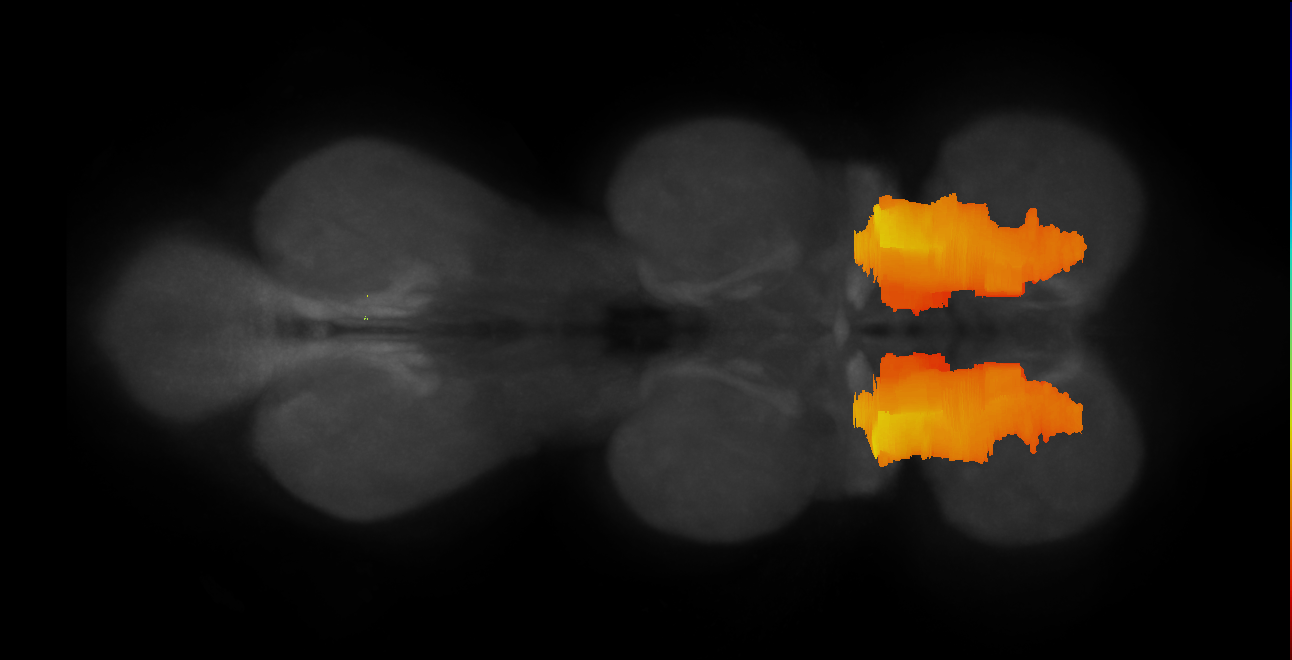

Imaging Technique
- computer graphic
Downloads
Image files aligned to wing tectulum on adult VNC, JRC2018VU:
Image files aligned to adult prothoracic neuromere on adult VNC, JRC2018VU:
Image files aligned to neck tectulum on adult VNC, JRC2018VU:
Image files aligned to adult metathoracic neuromere on adult VNC, JRC2018VU:
Image files aligned to adult mesothoracic neuromere on adult VNC, JRC2018VU:
Image files aligned to lower tectulum on adult VNC, JRC2018VU:
Image files aligned to intermediate tectulum on adult VNC, JRC2018VU:
Image files aligned to ITD haltere tract on adult VNC, JRC2018VU:
Image files aligned to median dorsal abdominal tract on adult VNC, JRC2018VU:
Image files aligned to dorsal lateral tract on adult VNC, JRC2018VU:
References
3 - Adult VNS neuropils (Court2017) [Court2017]
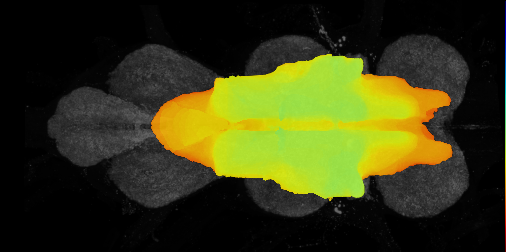
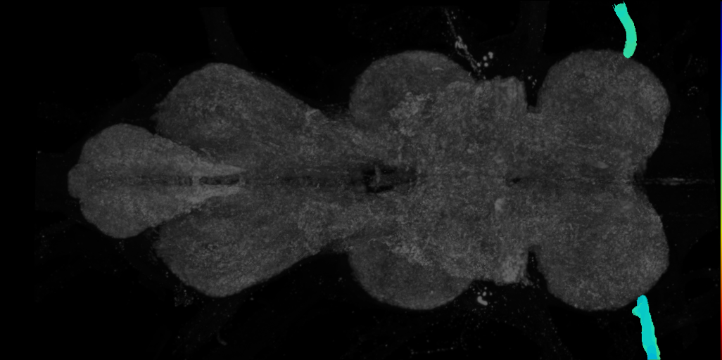
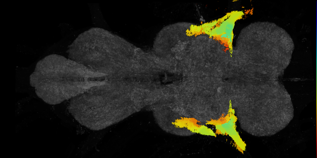
Imaging Technique
- computer graphic
- confocal microscopy
Downloads
Image files aligned to tectulum on adult VNS template, Court2018:
Image files aligned to accessory prothoracic nerve on adult VNS template, Court2018:
Image files aligned to anterior dorsal mesothoracic nerve on adult VNS template, Court2018:
Image files aligned to metathoracic leg nerve on adult VNS template, Court2018:
Image files aligned to abdominal segment 3 nerve on adult VNS template, Court2018:
Image files aligned to abdominal nerve trunk on adult VNS template, Court2018:
Image files aligned to adult abdominal neuromere on adult VNS template, Court2018:
Image files aligned to cervical connective on adult VNS template, Court2018:
Image files aligned to adult prothoracic neuromere on adult VNS template, Court2018:
Image files aligned to adult mesothoracic neuromere on adult VNS template, Court2018:
References
4 - AMMC local and projection neurons (Matsuo2016) [Matsuo2016]


Imaging Technique
- confocal microscopy
Downloads
Image files aligned to Db1:
Image files aligned to Bi7:
Image files aligned to Bi6:
Image files aligned to Bi2:
Image files aligned to Bb4:
Image files aligned to Db1:
Image files aligned to Bi7:
Image files aligned to Bi6:
Image files aligned to Bi2:
Image files aligned to Bb4:
References
5 - banc [Bates2025]
banc
ID: Bates2025
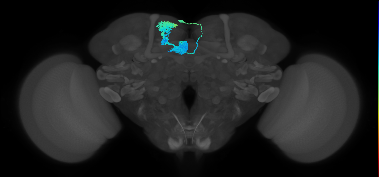
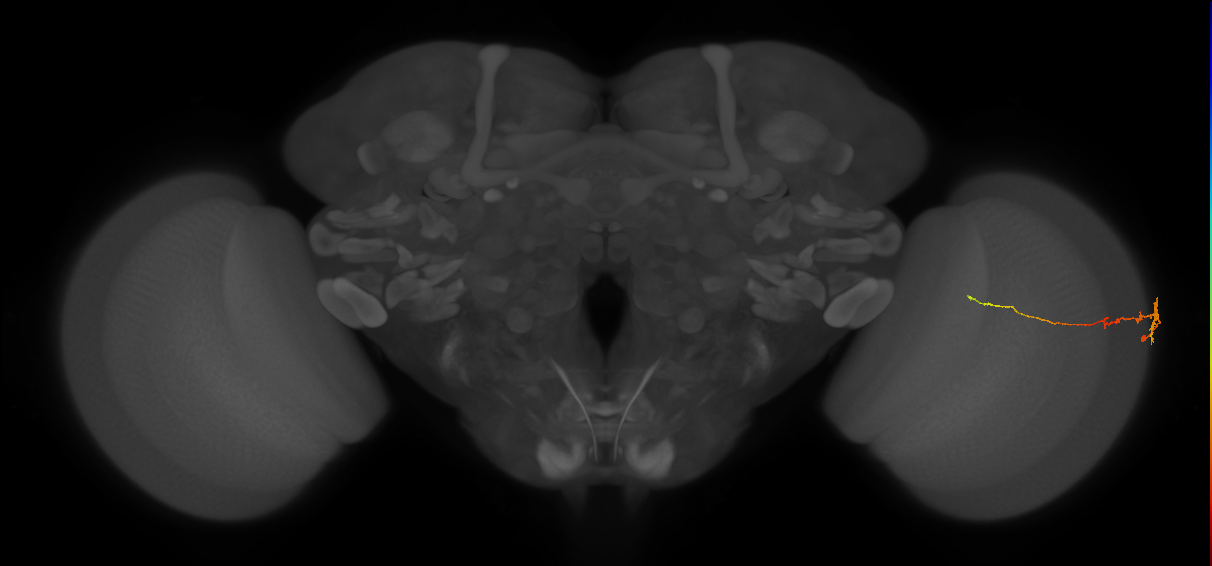
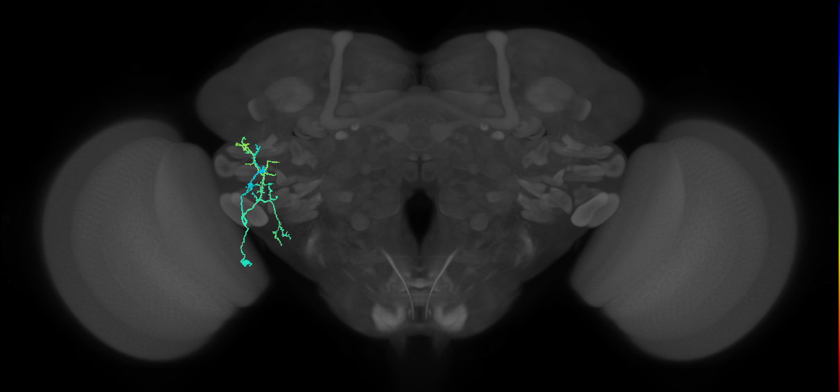
Neurons from Brain and Nerve Cord (BANC) - Bates et al., 2025. Includes neuron skeletons, meshes and their synaptic connectivity.
Open in VFB 3D Browser →Imaging Technique
- transmission electron microscopy (TEM)
Downloads
Image files aligned to BANC_626:720575941441821275:
Image files aligned to BANC_626:720575941431698529:
Image files aligned to BANC_626:720575941469997021:
Image files aligned to BANC_626:720575941524833538:
Image files aligned to BANC_626:720575941546887092:
Image files aligned to BANC_626:720575941561815720:
Image files aligned to BANC_626:720575941637721205:
Image files aligned to BANC_626:720575941479117698:
Image files aligned to BANC_626:720575941637721205:
Image files aligned to BANC_626:720575941659414096:
List all available images of banc (10 total)
| Thumbnail | Name | Tags |
|---|---|---|
| BANC_626:720575941441821275 | Nervous_system, Adult |
| BANC_626:720575941469997021 | Nervous_system, Adult |
 | BANC_626:720575941479117698 | Nervous_system, Adult |
 | BANC_626:720575941637721205 | Nervous_system, Adult |
 | BANC_626:720575941637721205 | Nervous_system, Adult |
Images in dataset banc (89045 total)
| Thumbnail | Name | Tags |
|---|---|---|
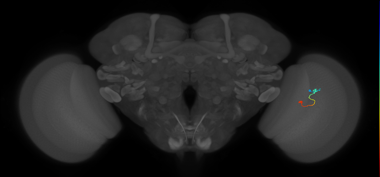 | [BANC_626:720575941391648406](/reports/VFB_00106ejz) | secondary_neuron, Nervous_system, Visual_system, Adult, Cholinergic |
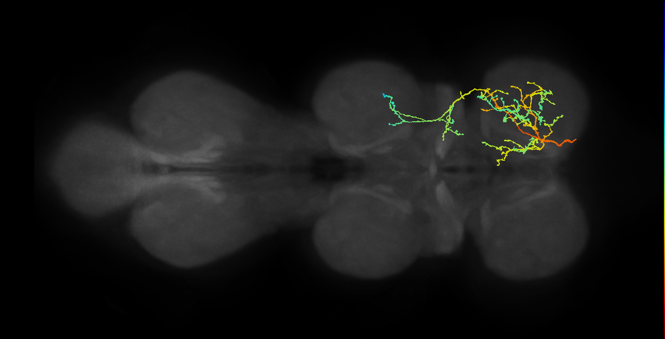 | [BANC_626:720575941432269271](/reports/VFB_00106ata) | Nervous_system, primary_neuron, Adult, Cholinergic |
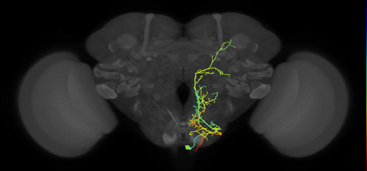 | [BANC_626:720575941432269271](/reports/VFB_00106ata) | Nervous_system, primary_neuron, Adult, Cholinergic |
 | [BANC_626:720575941463627533](/reports/VFB_001069v3) | Adult, Cholinergic, lineage_AOTUv4_DALcl2 |
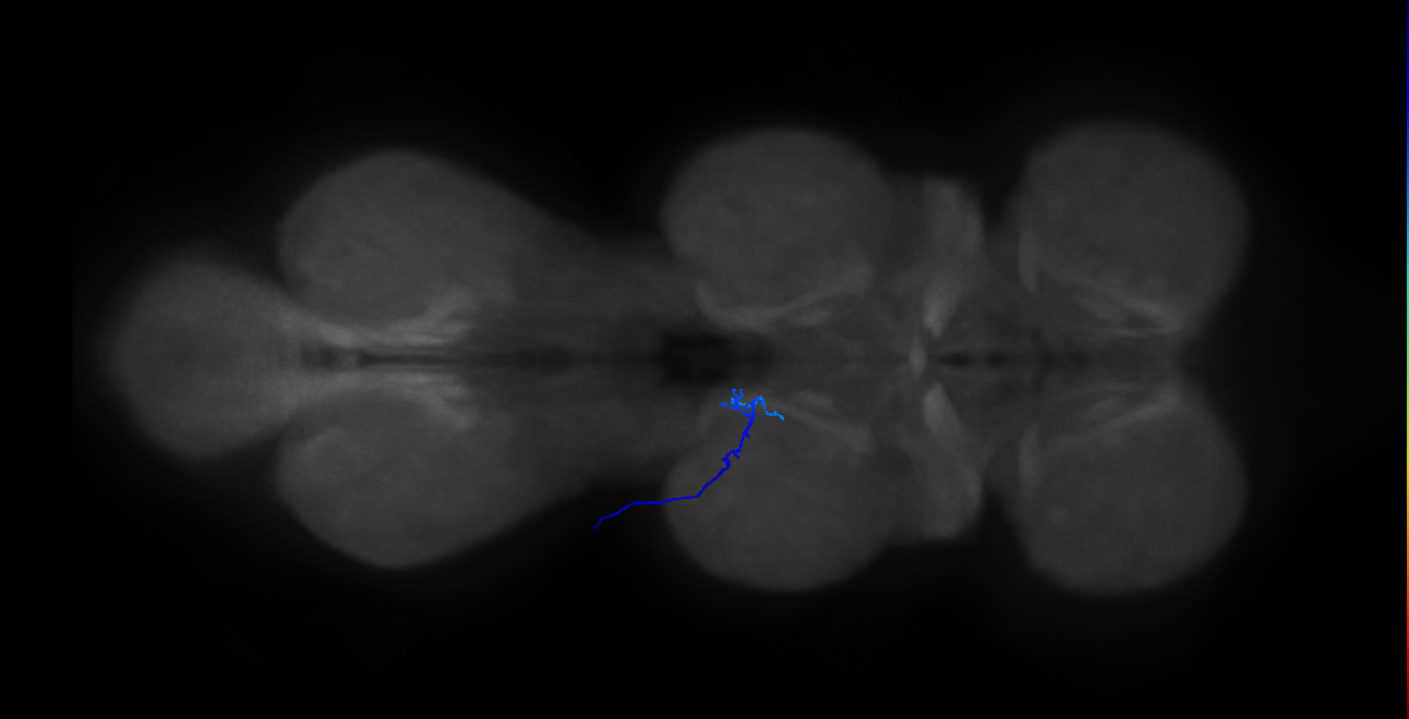 | [BANC_626:720575941451828807](/reports/VFB_001067c0) | Nervous_system, Sensory_neuron, Glutamatergic, Adult, Chemosensory_system |
 | [BANC_626:720575941514224633](/reports/VFB_00105sp4) | Nervous_system, Visual_system, Adult, GABAergic |
 | [BANC_626:720575941535965923](/reports/VFB_00105jxs) | secondary_neuron, Nervous_system, Visual_system, Adult, Cholinergic |
 | [BANC_626:720575941571781448](/reports/VFB_00105f3y) | Nervous_system, Adult, Visual_system, Cholinergic |
View all 89045 results in VFB →
References
6 - Biomechanical origins of proprioceptive maps in the Drosophila leg [Mamiya2022]


Biomechanical origins of proprioceptive maps in the Drosophila leg
ID: Mamiya2022
Biomechanical origins of proprioceptive maps in the Drosophila leg (Mamiya et al. 2022)
Open in VFB 3D Browser →Cross References
Imaging Technique
- X-ray computed tomography
Downloads
Image files aligned to FeCO lateral tendon (T1LEG:1688899):
Image files aligned to FeCO medial tendon (T1LEG:1688887):
Image files aligned to arculum outline (T1LEG:1688874):
Image files aligned to arculum midline (T1LEG:1688860):
References
7 - BrainName neuropils and tracts - Ito half-brain [BrainName_Ito_half_brain]
BrainName neuropils and tracts - Ito half-brain
ID: BrainName_Ito_half_brain
Imaging Technique
- computer graphic
Downloads
Image files aligned to posterior posteriolateral protocerebral commissure on adult brain template Ito2014:
Image files aligned to posterior lateral fascicle on adult brain template Ito2014:
Image files aligned to adult gnathal ganglion on adult brain template Ito2014:
Image files aligned to posterior ventrolateral protocerebrum on adult brain template Ito2014:
Image files aligned to adult antennal lobe on adult brain template Ito2014:
Image files aligned to antler on adult brain template Ito2014:
Image files aligned to inferior bridge on adult brain template Ito2014:
Image files aligned to lateral accessory lobe on adult brain template Ito2014:
Image files aligned to protocerebral bridge on adult brain template Ito2014:
Image files aligned to nodulus on adult brain template Ito2014:
List all available images of BrainName neuropils and tracts - Ito half-brain (10 total)
| Thumbnail | Name | Tags |
|---|---|---|
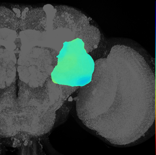
 | adult antennal lobe on adult brain template Ito2014 | Nervous_system, Adult, Synaptic_neuropil_domain |
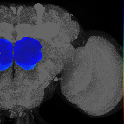
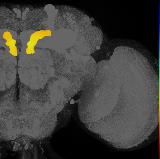 | antler on adult brain template Ito2014 | Nervous_system, Adult, Synaptic_neuropil_domain |
| posterior lateral fascicle on adult brain template Ito2014 | Nervous_system, Adult |
| posterior posteriolateral protocerebral commissure on adult brain template Ito2014 | Nervous_system, Adult |
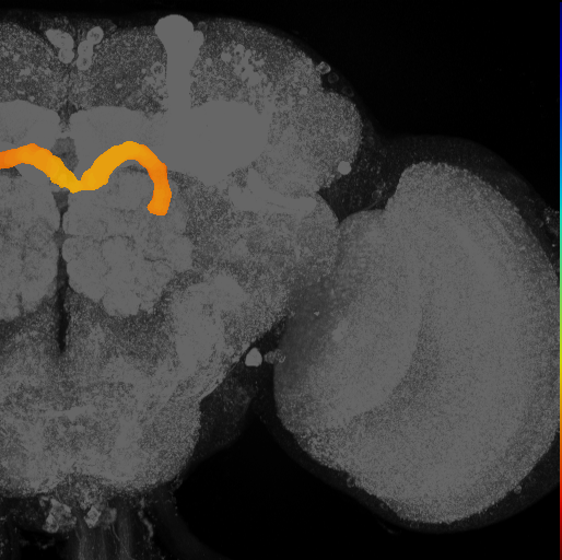 | protocerebral bridge on adult brain template Ito2014 | Nervous_system, Adult, Synaptic_neuropil_domain |
Images in dataset BrainName neuropils and tracts - Ito half-brain (75 total)
| Thumbnail | Name | Tags |
|---|---|---|
 | [posterior posteriolateral protocerebral commissure on adult brain template Ito2014](/reports/VFB_00030857) | Nervous_system, Adult, Neuron_projection_bundle |
 | [posterior lateral fascicle on adult brain template Ito2014](/reports/VFB_00030855) | Nervous_system, Adult, Neuron_projection_bundle |
 | [adult gnathal ganglion on adult brain template Ito2014](/reports/VFB_00030833) | Nervous_system, Adult, Ganglion |
 | [posterior ventrolateral protocerebrum on adult brain template Ito2014](/reports/VFB_00030816) | Nervous_system, Adult, Synaptic_neuropil_domain |
 | [adult antennal lobe on adult brain template Ito2014](/reports/VFB_00030809) | Nervous_system, Adult, Synaptic_neuropil_domain |
 | [antler on adult brain template Ito2014](/reports/VFB_00030800) | Nervous_system, Adult, Synaptic_neuropil_domain |
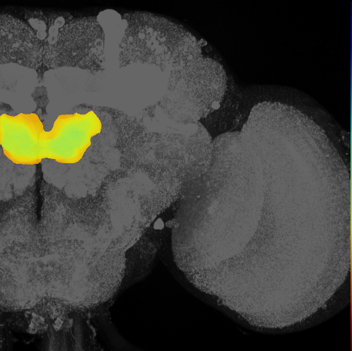 | [inferior bridge on adult brain template Ito2014](/reports/VFB_00030799) | Nervous_system, Adult, Synaptic_neuropil_domain |
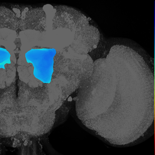 | [lateral accessory lobe on adult brain template Ito2014](/reports/VFB_00030793) | Nervous_system, Adult, Synaptic_neuropil_domain |
 | [protocerebral bridge on adult brain template Ito2014](/reports/VFB_00030791) | Nervous_system, Adult, Synaptic_neuropil_domain |
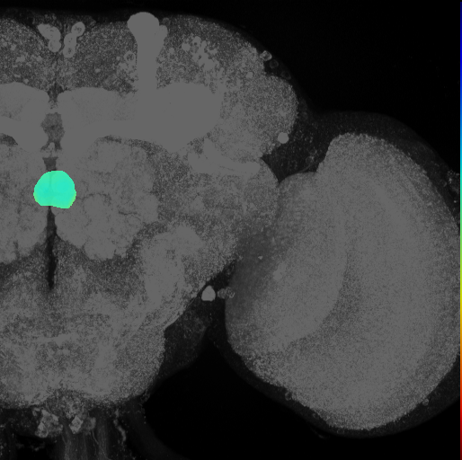 | [nodulus on adult brain template Ito2014](/reports/VFB_00030789) | Nervous_system, Adult, Synaptic_neuropil_domain |
References
8 - BrainName neuropils on adult brain JFRC2 (Jenett, Shinomya) [JenettShinomya_BrainName]


BrainName neuropils on adult brain JFRC2 (Jenett, Shinomya)
ID: JenettShinomya_BrainName
Imaging Technique
- computer graphic
Downloads
Image files aligned to vertical lobe of adult mushroom body on adult brain template JFRC2:
Image files aligned to lobe system of adult mushroom body on adult brain template JFRC2:
Image files aligned to lateral complex on adult brain template JFRC2:
Image files aligned to adult subesophageal zone on adult brain template JFRC2:
Image files aligned to SIP on adult brain template JFRC2:
Image files aligned to medulla on adult brain template JFRC2:
Image files aligned to superior clamp on adult brain template JFRC2:
Image files aligned to ellipsoid body on adult brain template JFRC2:
Image files aligned to anterior ventrolateral protocerebrum on adult brain template JFRC2:
Image files aligned to epaulette on adult brain template JFRC2:
References
9 - BrainTrap lines (Knowles-Barley2010) [Knowles_Barley2010]


Cross References
Imaging Technique
- confocal microscopy
References
10 - central brain neurons by lineage, Lee2020 [Lee_Lineage2020]
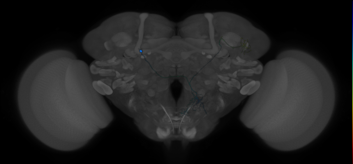
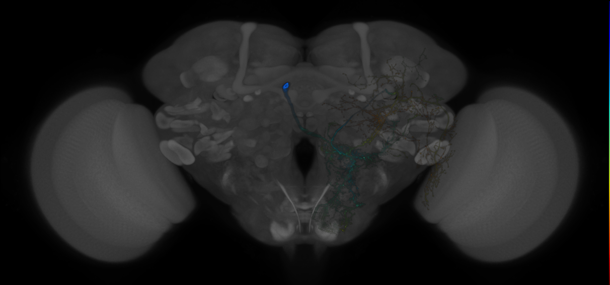
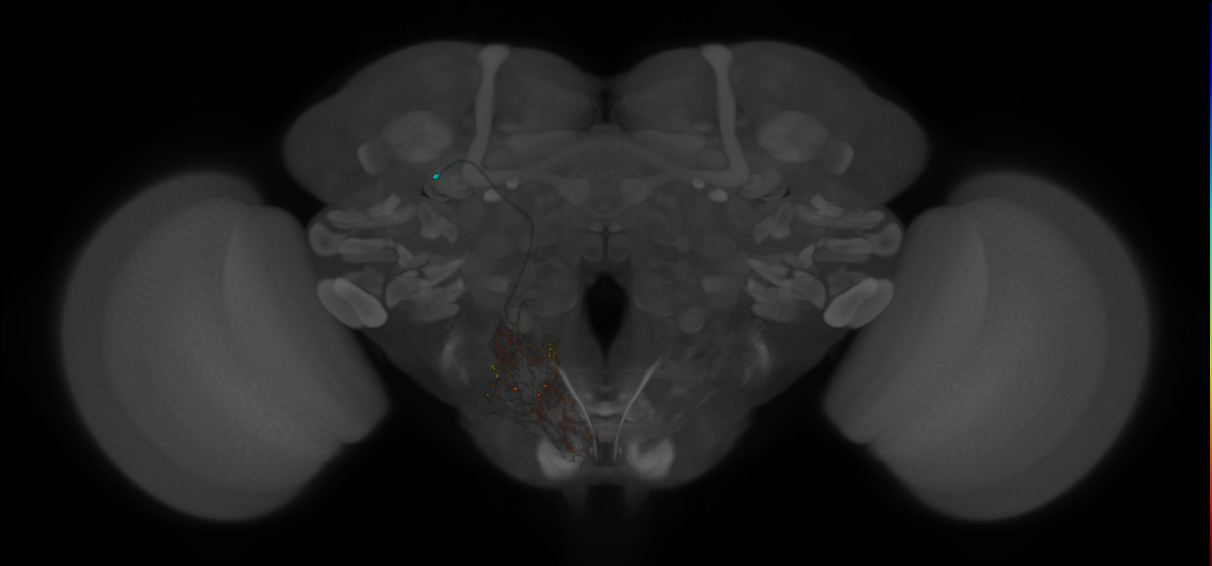
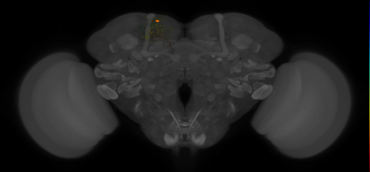
central brain neurons by lineage, Lee2020
ID: Lee_Lineage2020
A catalogue of single neuron images covering 18 central brain lineages.
Open in VFB 3D Browser →Imaging Technique
- confocal microscopy
Downloads
Image files aligned to CREa1B_P07(SLP)_0_2018U:
Image files aligned to CREa1B_P01(LO)_0_2018U:
Image files aligned to WEDd1_S02(PS)_0_2018U:
Image files aligned to SMPp&v1A_S03(SP)_0_2018U:
Image files aligned to SMPp&v1A_H06(_CL)_0_2018U:
Image files aligned to LALv1B_H01(_IPS)_0_2018U:
Image files aligned to AOTUv4A_M01(FB)_0_2018U:
Image files aligned to AOTUv3B_P01(LAL)_0_2018U:
Image files aligned to AOTUv3A_H10(PS)_0_2018U:
Image files aligned to AOTUv1B_S13(PS)_0_2018U:
References
11 - Comparative Connectomics Reveals How Partner Identity, Location, and Activity Specify Synaptic Connectivity in Drosophila (Valdes-Aleman et al. 2021) [Valdes_Aleman2021]


Comparative Connectomics Reveals How Partner Identity, Location, and Activity Specify Synaptic Connectivity in Drosophila (Valdes-Aleman et al. 2021)
ID: Valdes_Aleman2021
L1 EM reconstructed neurons from Valdes-Aleman et al., 2021
Open in VFB 3D Browser →Cross References
Imaging Technique
- transmission electron microscopy (TEM)
Downloads
Image files aligned to Drunken-1 a1l (L1EM:6207232):
Image files aligned to Griddle-1 a1r (L1EM:4824878):
Image files aligned to Drunken-1 a1r (L1EM:4096366):
Image files aligned to Basin3_a1 (L1EM:10179501):
Image files aligned to chos_lch5-3_a1 (L1EM:8031834):
Image files aligned to chos_lch5-1_a1 (L1EM:7764785):
Image files aligned to Basin3_a1 (L1EM:3091943):
Image files aligned to Basin2_a1 (L1EM:3041612):
Image files aligned to chos_lch5-24-1_a1 (L1EM:7577773):
Image files aligned to chos_lch5-24-2_a1 (L1EM:159280):
References
12 - Dickson lab VT line collection - VDRC images [Dickson_VT]


Cross References
Imaging Technique
- confocal microscopy
Downloads
Image files aligned to VT061192:
Image files aligned to VT055399:
Image files aligned to VDRC_VT044170_LexAGAD_attP40_1:
Image files aligned to VDRC_VT007843_LexAGAD_attP40_2:
Image files aligned to VT007378:
Image files aligned to VT061192:
Image files aligned to VT055399:
Image files aligned to VDRC_VT044170_LexAGAD_attP40_1:
Image files aligned to VDRC_VT007843_LexAGAD_attP40_2:
Image files aligned to VT007378:
References
13 - Dickson lab VT lines - FlyLight/Janelia images (2017) [Dickson2017]


Dickson lab VT lines - FlyLight/Janelia images (2017)
ID: Dickson2017
Cross References
Imaging Technique
- confocal microscopy
Downloads
Image files aligned to VT045983:
Image files aligned to VT038440:
Image files aligned to VT010258:
Image files aligned to VT045983:
Image files aligned to VT038440:
Image files aligned to VT024098:
Image files aligned to VT010258:
Image files aligned to VT064878:
Image files aligned to VT039567:
Image files aligned to VT021367:
References
14 - EM FAFB Baltruschat et al 2021 [Baltruschat2021]
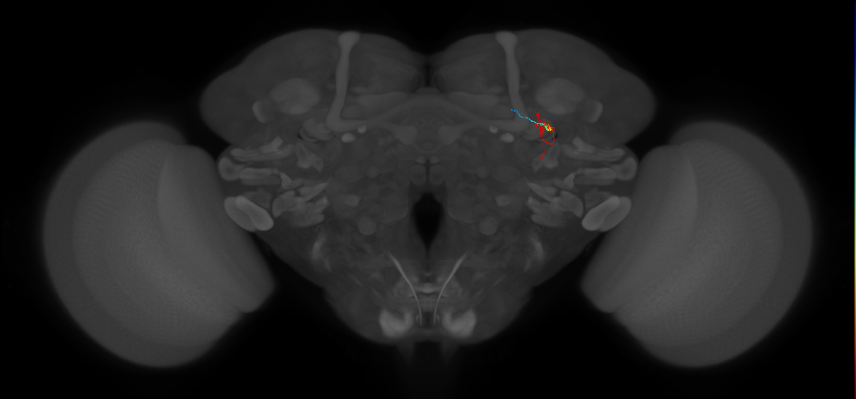
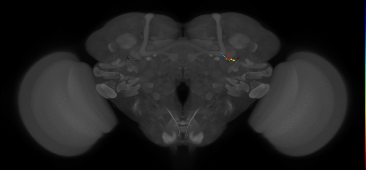
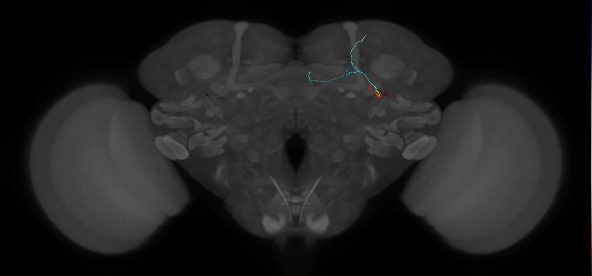
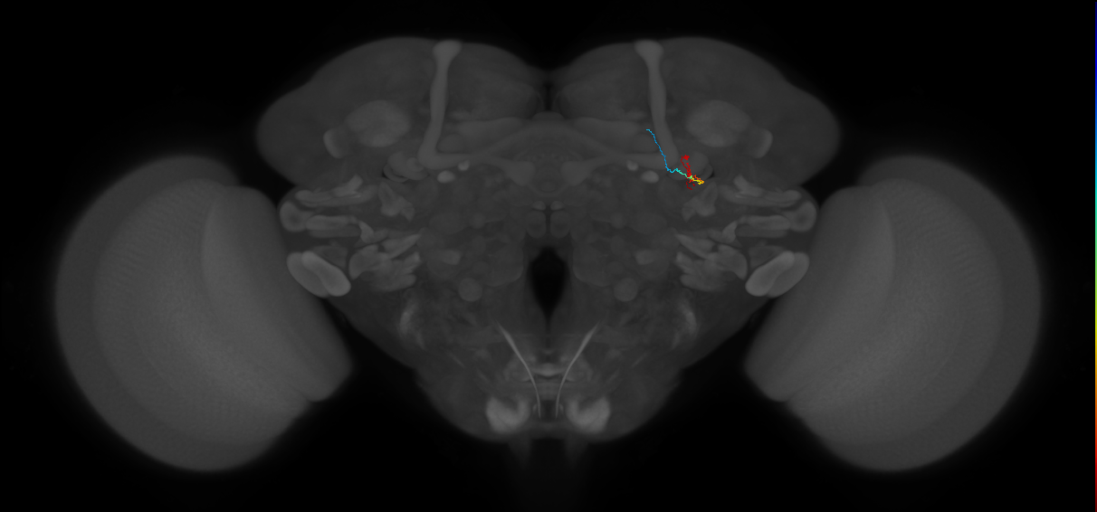
EM FAFB Baltruschat et al 2021
ID: Baltruschat2021
FAFB EM reconstructed neurons from Baltruschat et al 2021
Open in VFB 3D Browser →Cross References
Imaging Technique
- transmission electron microscopy (TEM)
Downloads
Image files aligned to KC#1513-ym (FAFB:2718221):
Image files aligned to KC#1512-ym (FAFB:541622):
Image files aligned to KC#1509-a'b'm (FAFB:527842):
Image files aligned to KC#1510-ym (FAFB:245402):
Image files aligned to MB-C1#1 (FAFB:535060):
Image files aligned to putative MB-C1 32768 JMR (FAFB:60445):
Image files aligned to APL 203841 MR JSL (FAFB:203840):
Image files aligned to KC#559-y (FAFB:591301):
Image files aligned to KC#556-y (FAFB:535010):
Image files aligned to KC#379-y (FAFB:531058):
References
15 - EM FAFB Bates and Schlegel et al 2020 [BatesSchlegel2020]

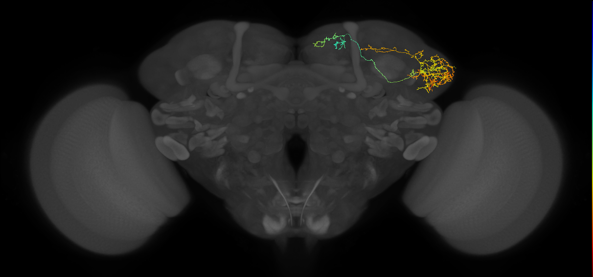
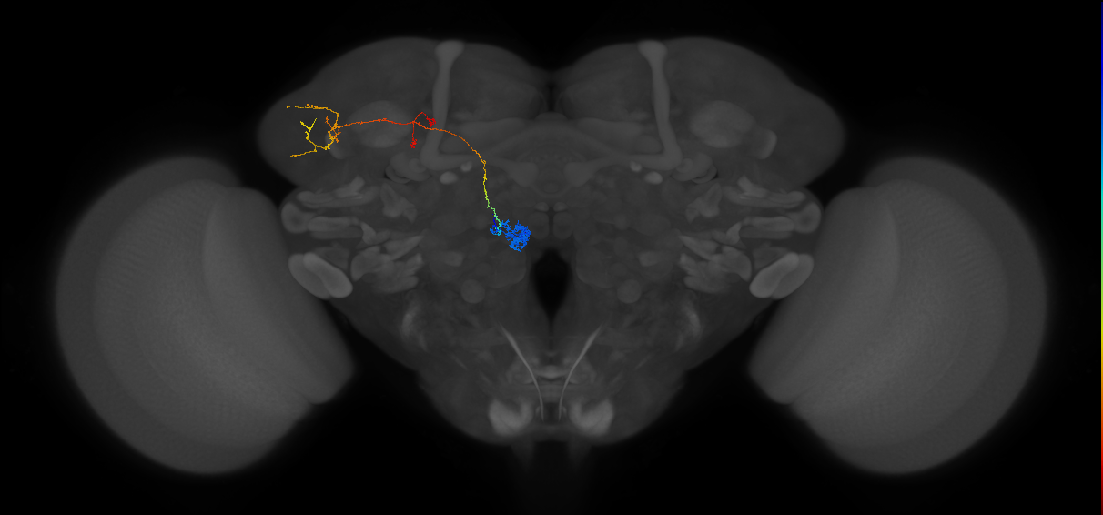
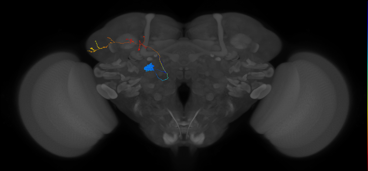
EM FAFB Bates and Schlegel et al 2020
ID: BatesSchlegel2020
FAFB EM reconstructed neurons from Bates and Schlegel et al 2020
Open in VFB 3D Browser →Cross References
Imaging Technique
- transmission electron microscopy (TEM)
Downloads
Image files aligned to LHPD3a2#1 (FAFB:4182038):
Image files aligned to LHAD1b2#7 (FAFB:3080225):
Image files aligned to Uniglomerular mALT VM7d adPN#L1 (FAFB:10533089):
Image files aligned to Uniglomerular mALT DL1 adPN#L2 (FAFB:5305037):
Image files aligned to LHAD1b2#3 (FAFB:5030649):
Image files aligned to aSP-g1b#1 (FAFB:2477473):
Image files aligned to Uniglomerular mALT DC3 adPN#L1 (FAFB:2474754):
Image files aligned to Multiglomerular mlALT vPN#R62 (FAFB:2182498):
Image files aligned to Multiglomerular mALT lvPN#R15 (FAFB:56983):
Image files aligned to Uniglomerular mALT VA1v adPN#R1 (FAFB:57246):
References
16 - EM FAFB Coates et al 2020 [Coates2020]
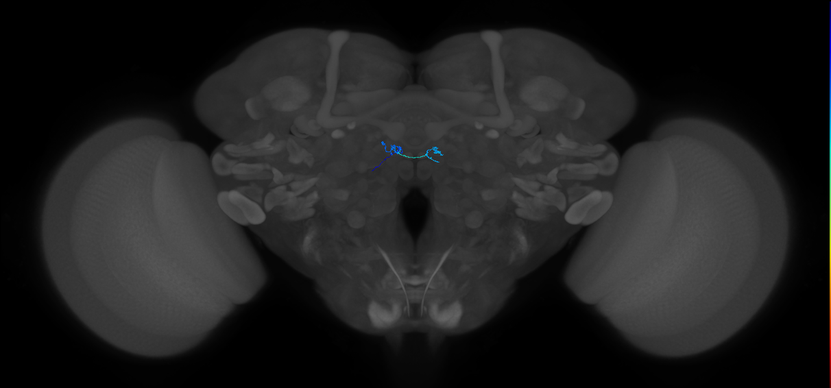
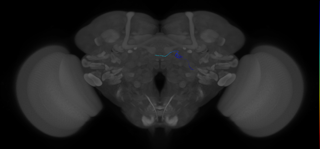
EM FAFB Coates et al 2020
ID: Coates2020
FAFB EM reconstructed neurons from Coates et al 2020
Open in VFB 3D Browser →Cross References
Imaging Technique
- transmission electron microscopy (TEM)
Downloads
Image files aligned to DM1 ORN #17 (FAFB:14545747):
Image files aligned to DA2 ORN #21 (FAFB:7490413):
Image files aligned to DA2 ORN #15 (FAFB:7349870):
Image files aligned to DA2 ORN #13 (FAFB:7349832):
Image files aligned to Patchy LN #9 (FAFB:7298540):
Image files aligned to DM1 ORN #11 (FAFB:7179380):
Image files aligned to Patchy LN #6 (FAFB:6190457):
Image files aligned to DA2 ORN #7 (FAFB:5007809):
Image files aligned to DM1 ORN #6 (FAFB:4079928):
Image files aligned to Dense ABAF LN#2 (FAFB:2891306):
References
17 - EM FAFB Dolan and Belliart-Guerin et al. 2018 [Dolan2018]


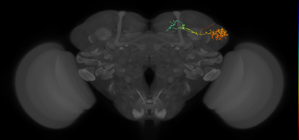
EM FAFB Dolan and Belliart-Guerin et al. 2018
ID: Dolan2018
FAFB EM reconstructed neurons from Dolan and Belliart-Guerin et al. 2018
Open in VFB 3D Browser →Cross References
Imaging Technique
- transmission electron microscopy (TEM)
Downloads
Image files aligned to MBON-a2sc_L (FAFB:1420974):
Image files aligned to MBON-a2sc_R (FAFB:346114):
Image files aligned to MBON-ap2_R (FAFB:1540941):
Image files aligned to LHPD2a1#5 (FAFB:11547665):
Image files aligned to LHPD2b1#2 (FAFB:3345012):
Image files aligned to LHPD2a1#2 (FAFB:2205218):
Image files aligned to LHPD2b1#1 (FAFB:1606113):
Image files aligned to LHPD2a1#4 (FAFB:1454234):
Image files aligned to LHPD2a1#3 (FAFB:1415893):
Image files aligned to LHPD2a1#1 (FAFB:1299700):
References
18 - EM FAFB Dolan et al. 2019 [FafbDolan2019]
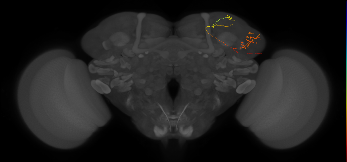
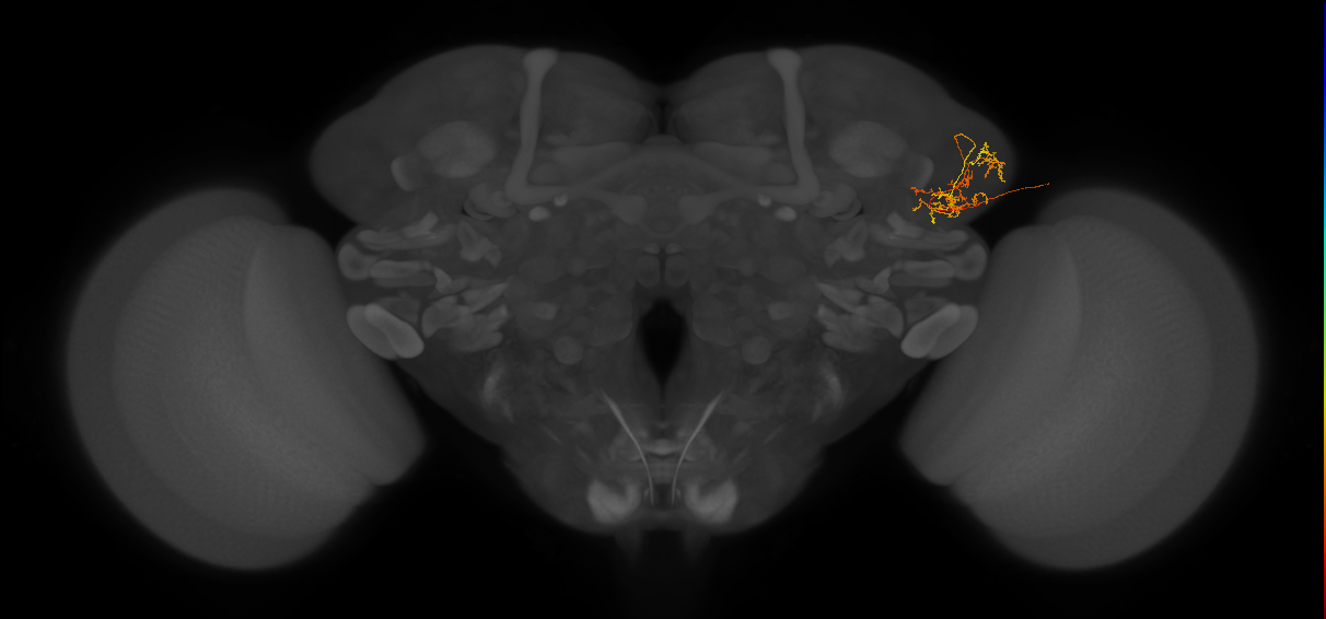


EM FAFB Dolan et al. 2019
ID: FafbDolan2019
FAFB EM reconstructed neurons from Dolan et al., 2019
Open in VFB 3D Browser →Cross References
Imaging Technique
- transmission electron microscopy (TEM)
Downloads
Image files aligned to LHPV5d1#2 (FAFB:11524200):
Image files aligned to LHPV4a2#2 (FAFB:4044614):
Image files aligned to VES002#1 (FAFB:4017748):
Image files aligned to LHPV4a7#1 (FAFB:3367291):
Image files aligned to LHPV6a3#2 (FAFB:2130631):
Image files aligned to LHAV4a4#2 (FAFB:1853423):
Image files aligned to WEDPN2B#2 (FAFB:9249174):
Image files aligned to LHAV6a1#1 (FAFB:1415926):
Image files aligned to aSP-g1b#1 (FAFB:2477473):
Image files aligned to MBON-Calyx_R (FAFB:30591):
References
19 - EM FAFB Dombrovski et al 2023 [Dombrovski2023]
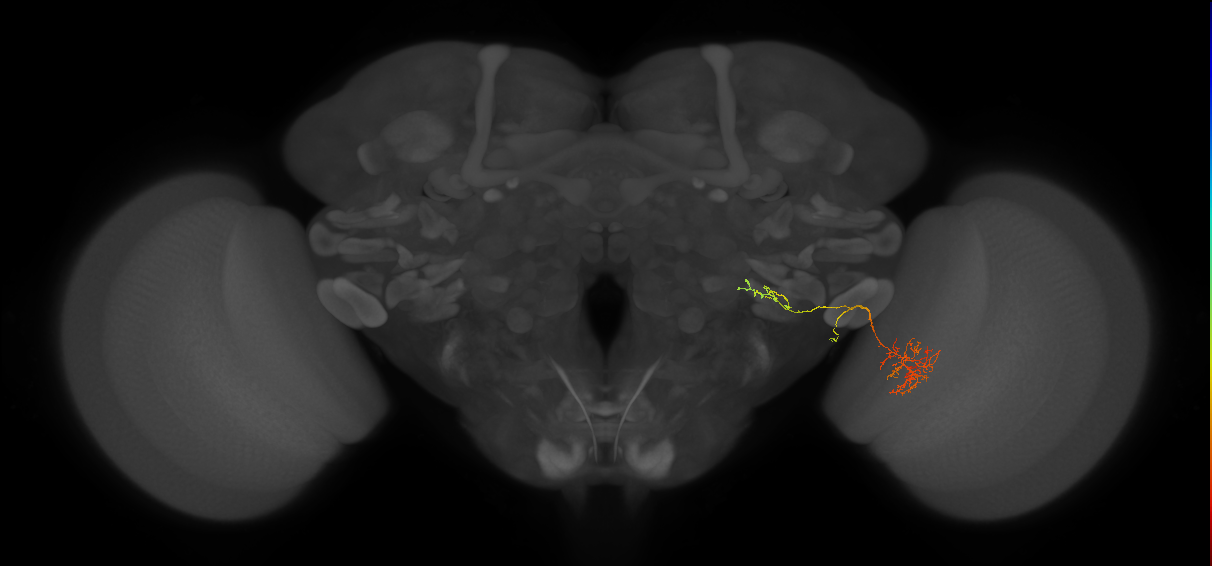
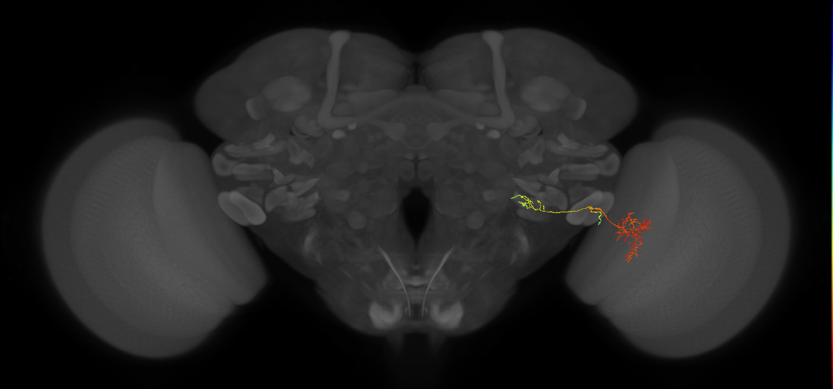
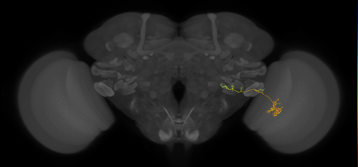
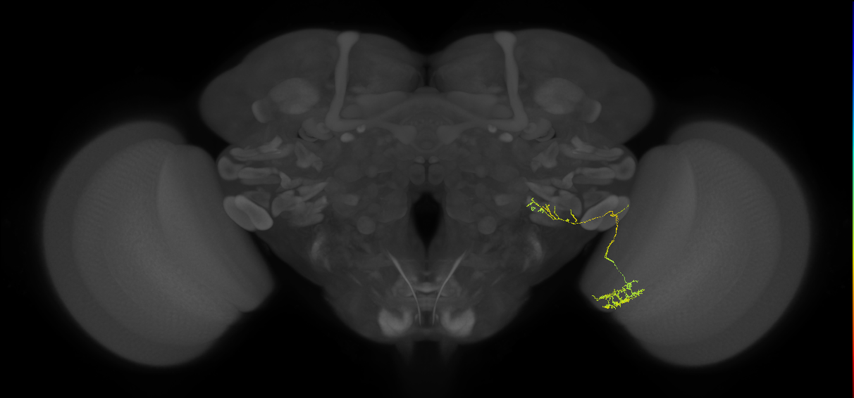
EM FAFB Dombrovski et al 2023
ID: Dombrovski2023
FAFB EM reconstructed neurons from Dombrovski et al., 2023
Open in VFB 3D Browser →Imaging Technique
- transmission electron microscopy (TEM)
Downloads
Image files aligned to LC4_R#39 (FAFB:1325197):
Image files aligned to LC4_R#37 (FAFB:1324489):
Image files aligned to LC4_R#32 (FAFB:1108134):
Image files aligned to LC4_R#29 (FAFB:1077372):
Image files aligned to LC4_R#25 (FAFB:1060322):
Image files aligned to LC4_R#13 (FAFB:872672):
Image files aligned to LC4_R#11 (FAFB:867557):
Image files aligned to LC4_R#6 (FAFB:308338):
Image files aligned to LC4_R#5 (FAFB:292771):
Image files aligned to LC4_R#4 (FAFB:292337):
20 - EM FAFB Engert et al. 2022 [Engert2022]
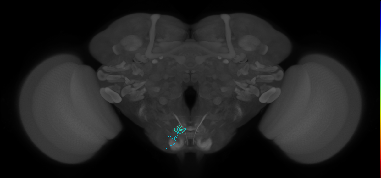

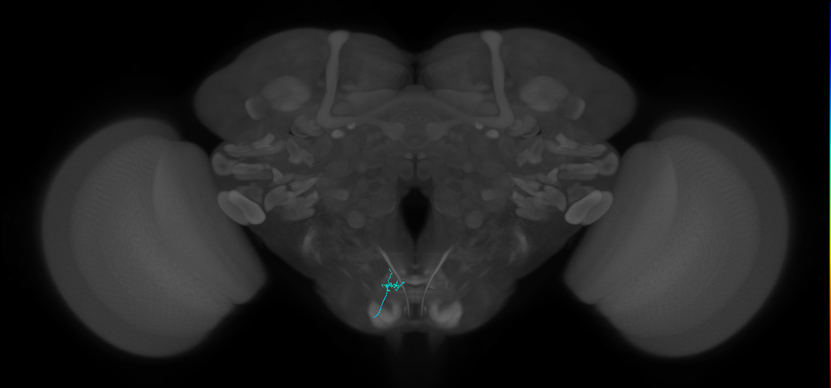
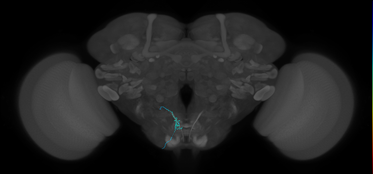
EM FAFB Engert et al. 2022
ID: Engert2022
FAFB EM reconstructed neurons from Engert et al 2022
Open in VFB 3D Browser →Cross References
Imaging Technique
- transmission electron microscopy (TEM)
Downloads
Image files aligned to GRN L4#9 (FAFB:16149095):
Image files aligned to GRN R6#23 (FAFB:14947958):
Image files aligned to GRN L4#5 (FAFB:14444228):
Image files aligned to GRN L7#4 (FAFB:11461075):
Image files aligned to GRN R3#8 (FAFB:8210748):
Image files aligned to GRN R1#1 (FAFB:7759923):
Image files aligned to GRN R2#2 (FAFB:7757554):
Image files aligned to GRN R6#6 (FAFB:7716995):
Image files aligned to GRN R4#4 (FAFB:7456677):
Image files aligned to GRN R4#1 (FAFB:7349219):
References
21 - EM FAFB Erginkaya et al 2025 [Erginkaya2025]
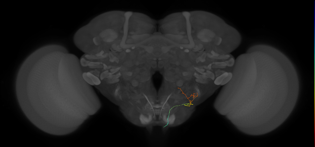
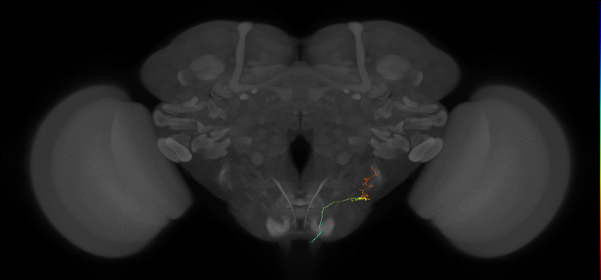
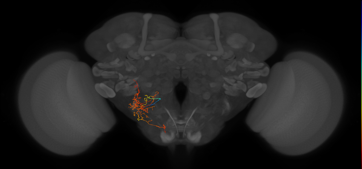
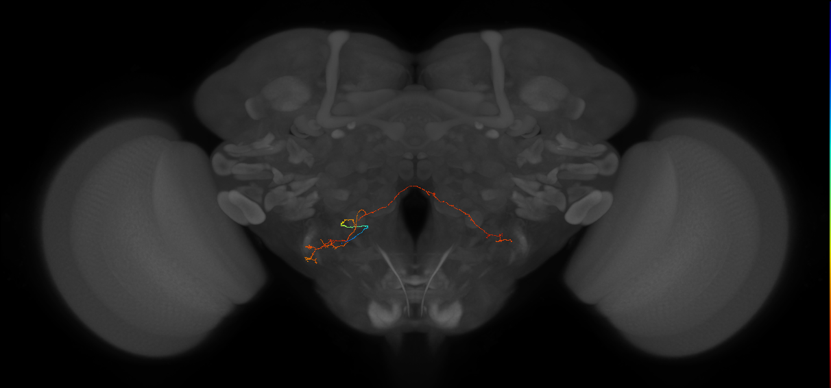
EM FAFB Erginkaya et al 2025
ID: Erginkaya2025
FAFB EM reconstructed neurons from Erginkaya et al 2025
Open in VFB 3D Browser →Cross References
Imaging Technique
- transmission electron microscopy (TEM)
Downloads
Image files aligned to right H2rn#5 (FAFB:11956980):
Image files aligned to right H2rn#4 (FAFB:11930921):
Image files aligned to left uLPTCrn#6 (FAFB:9157788):
Image files aligned to left bLPTCrn#3 (FAFB:9157627):
Image files aligned to right H2rn#1 (FAFB:8178073):
Image files aligned to left uLPTCrn#3 (FAFB:4577828):
Image files aligned to left uLPTCrn#1 (FAFB:3768475):
Image files aligned to right HSE (FAFB:827034):
Image files aligned to right VS6 (FAFB:804539):
Image files aligned to right cLPTCrn#5 (FAFB:411565):
References
22 - EM FAFB Felsenberg et al. 2018 [Felsenberg2018]
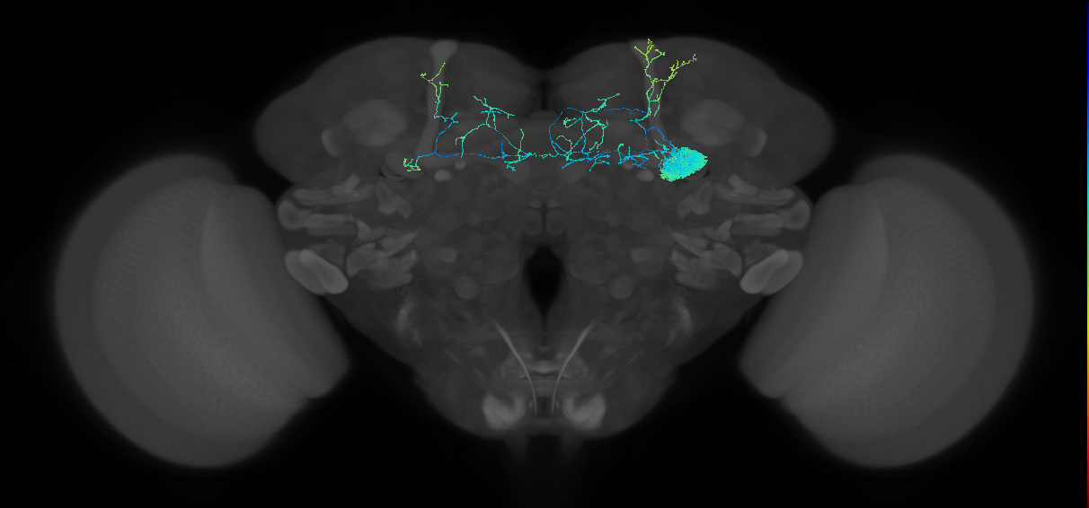
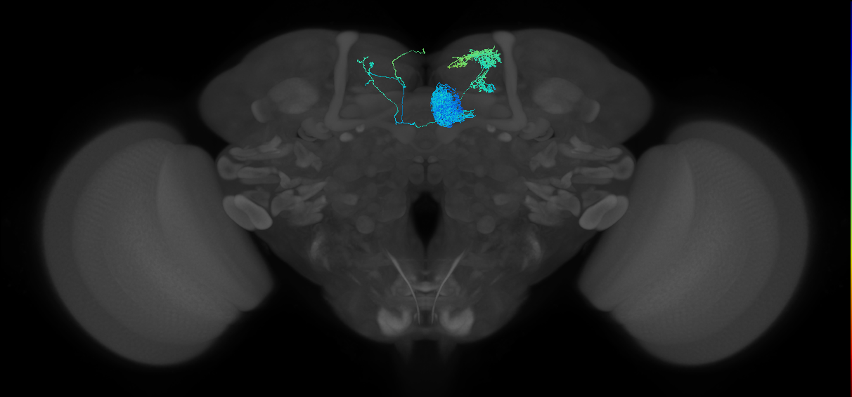

EM FAFB Felsenberg et al. 2018
ID: Felsenberg2018
FAFB EM reconstructed neurons from Felsenberg et al 2018
Open in VFB 3D Browser →Cross References
Imaging Technique
- transmission electron microscopy (TEM)
Downloads
Image files aligned to MBON-y1pedc>a/B_R (FAFB:2333007):
Image files aligned to MBON-y5B'2a_R (FAFB:2109445):
Image files aligned to MBON-B'2mp_R (FAFB:4291899):
References
23 - EM FAFB Gorko et al 2024 [Gorko2024]


EM FAFB Gorko et al 2024
ID: Gorko2024
FAFB EM reconstructed neurons from Gorko et al 2024
Open in VFB 3D Browser →Cross References
Imaging Technique
- transmission electron microscopy (TEM)
Downloads
Image files aligned to CvN7_NMN (FAFB:1337777):
Image files aligned to CvN6_NMN (FAFB:1167858):
References
24 - EM FAFB Hampel and Eichler et al 2020 [HampelEichler2020]

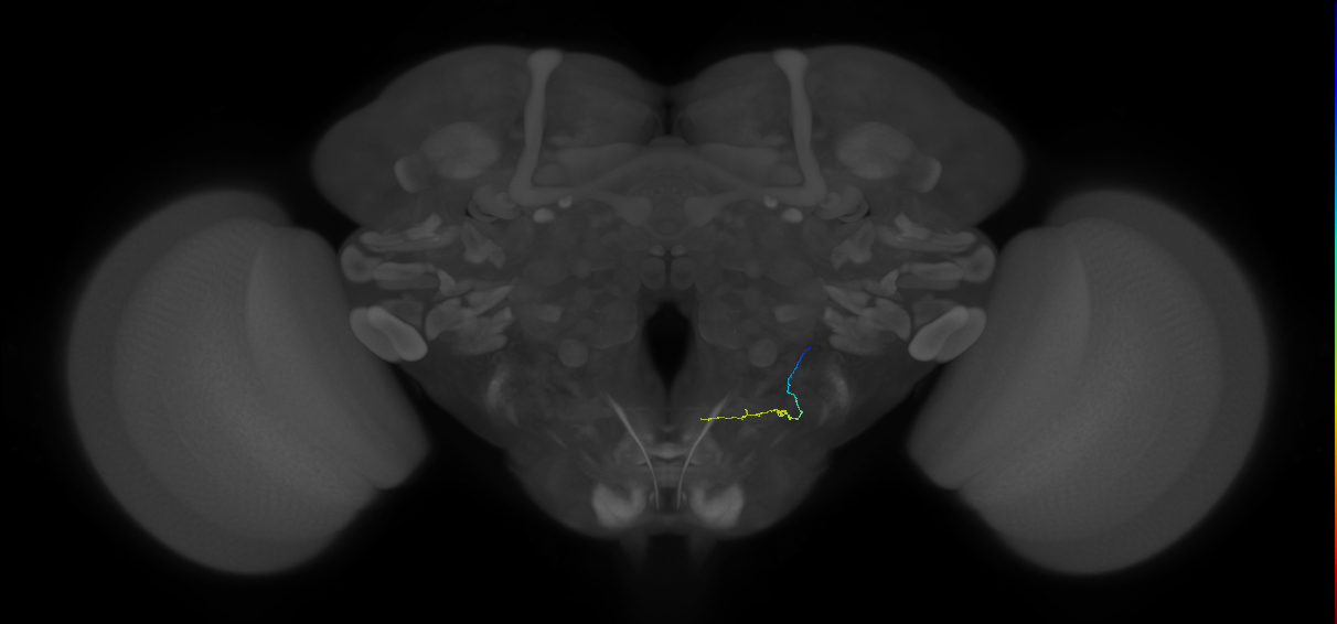

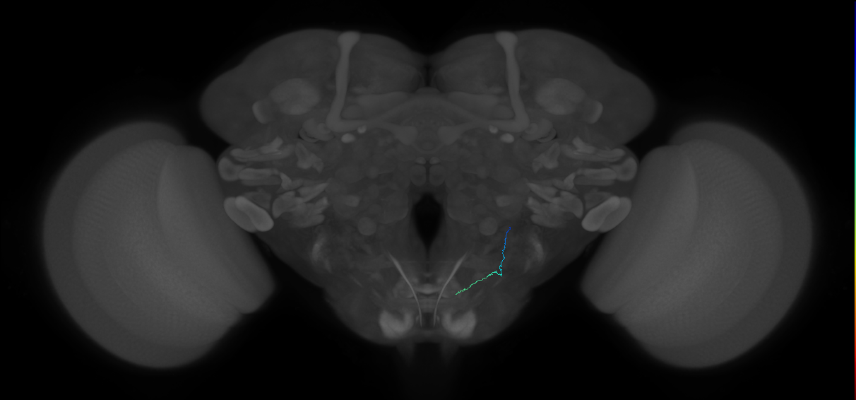
EM FAFB Hampel and Eichler et al 2020
ID: HampelEichler2020
FAFB EM reconstructed neurons from Hampel and Eichler et al 2020
Open in VFB 3D Browser →Cross References
Imaging Technique
- transmission electron microscopy (TEM)
Downloads
Image files aligned to JO-EVP#2 (FAFB:13587125):
Image files aligned to JO-EDM#10 (FAFB:13251040):
Image files aligned to JO-FVA#25 (FAFB:11369216):
Image files aligned to JO-FVA#24 (FAFB:11356453):
Image files aligned to JO-FDP#4 (FAFB:11090188):
Image files aligned to JO-FDP#2 (FAFB:10896629):
Image files aligned to JO-FVA#26 (FAFB:10771158):
Image files aligned to JO-CA#1 (FAFB:4524559):
Image files aligned to JO-EV#2 (FAFB:1600551):
Image files aligned to JO-EVP#1 (FAFB:1181773):
References
25 - EM FAFB Kim et al 2020 [Kim2020]
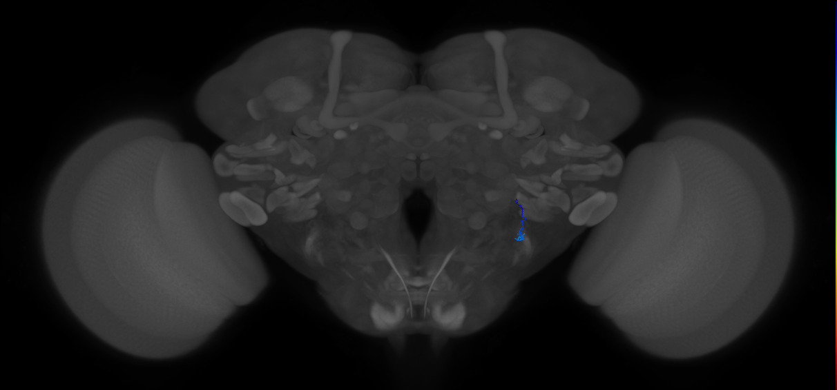


EM FAFB Kim et al 2020
ID: Kim2020
FAFB EM reconstructed neurons from Kim et al 2020
Open in VFB 3D Browser →Cross References
Imaging Technique
- transmission electron microscopy (TEM)
Downloads
Image files aligned to JO-B-10 (type-1) (FAFB:3674078):
Image files aligned to JO-B-21 (type-2) (FAFB:3642969):
Image files aligned to JO-A-41 (type-2) (FAFB:3529301):
Image files aligned to JO-A-40 (type-2) (FAFB:3517017):
Image files aligned to JO-A-36 (type-2) (FAFB:3488976):
Image files aligned to JO-A-3 (type-1) (FAFB:3449992):
Image files aligned to JO-A-27 (type-2) (FAFB:3446283):
Image files aligned to JO-B-23 (type-2) (FAFB:3402380):
Image files aligned to JO-B-25 (type-3) (FAFB:3387407):
Image files aligned to JO-B-36 (type-4) (FAFB:3164346):
References
26 - EM FAFB Kind et al. 2021 [Kind2021]
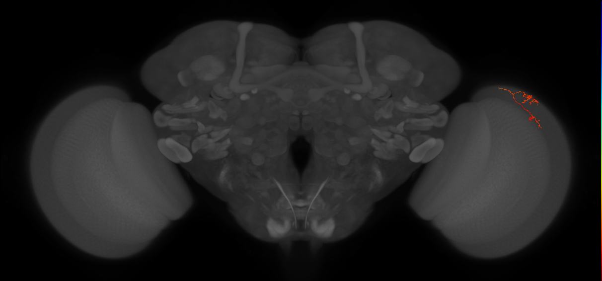
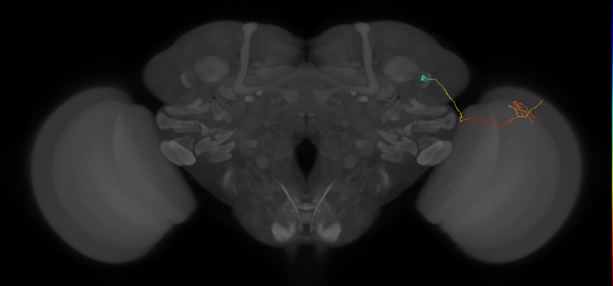
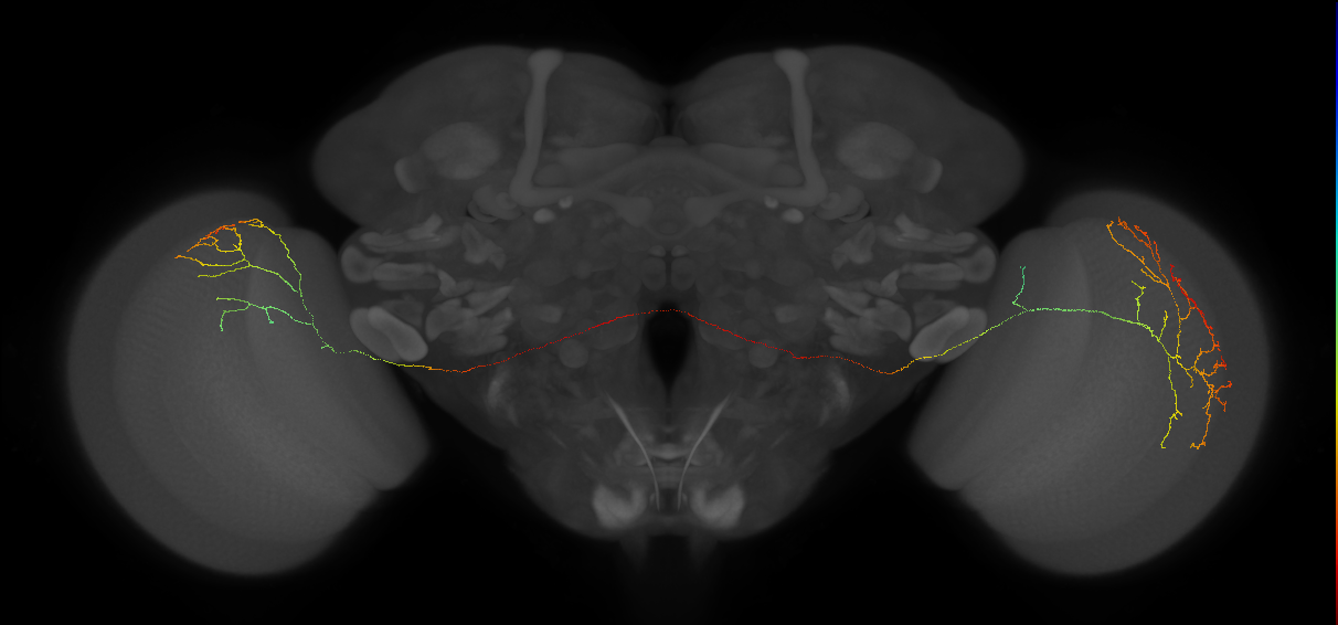
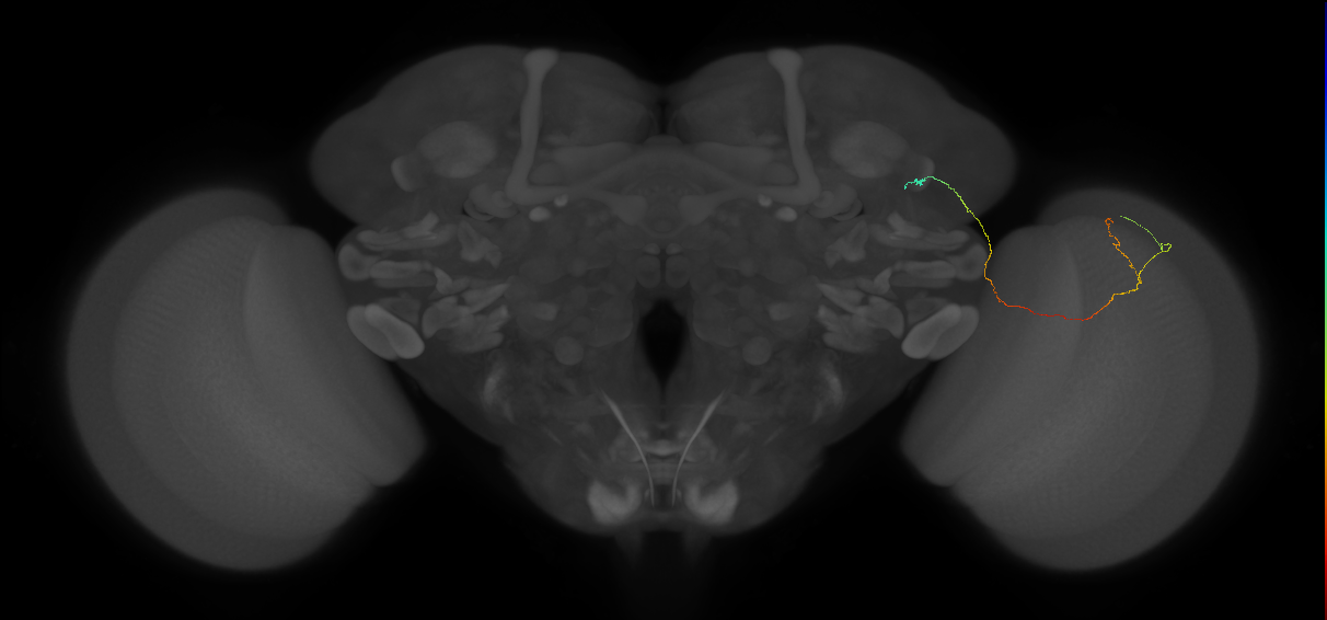
EM FAFB Kind et al. 2021
ID: Kind2021
FAFB EM reconstructed neurons from Kind et al., 2021
Open in VFB 3D Browser →Cross References
Imaging Technique
- transmission electron microscopy (TEM)
Downloads
Image files aligned to Dm9#11 (FAFB:14933802):
Image files aligned to MeTu#15 (FAFB:14726392):
Image files aligned to MeMe_DRA#2 (FAFB:11993543):
Image files aligned to MeTu_DRA#18 (FAFB:11993382):
Image files aligned to MeTu_DRA#24 (FAFB:11908743):
Image files aligned to Mti#1 (FAFB:11666155):
Image files aligned to C2#2 (FAFB:11472485):
Image files aligned to MeTu#9 (FAFB:10656250):
Image files aligned to R8_DRA#3 (FAFB:10190508):
Image files aligned to L1#2 (FAFB:10108985):
References
27 - EM FAFB Marin et al 2020 [Marin2020]


EM FAFB Marin et al 2020
ID: Marin2020
FAFB EM reconstructed neurons from Marin et al 2020
Open in VFB 3D Browser →Cross References
Imaging Technique
- transmission electron microscopy (TEM)
Downloads
Image files aligned to VP2 l2PN_L (FAFB:13154015):
Image files aligned to VP4RNl#11 (FAFB:12819401):
Image files aligned to VP2+VC5 l2PN_L (FAFB:10515783):
Image files aligned to VP1d+VP4 l2PN2_L (FAFB:9740174):
Image files aligned to VP1d+VP4 l2PN1_L (FAFB:9716626):
Image files aligned to VP5RNl#4 (FAFB:9612139):
Image files aligned to PD5g1#3_L (FAFB:8880131):
Image files aligned to VP1mRNr#3 (FAFB:4889524):
Image files aligned to Multiglomerular mALT lPN bilateral#R2 (FAFB:57434):
Image files aligned to Uniglomerular mALT VP2+ adPN#R1 (FAFB:57516):
References
28 - EM FAFB Morimoto et al 2020 [Morimoto2020]
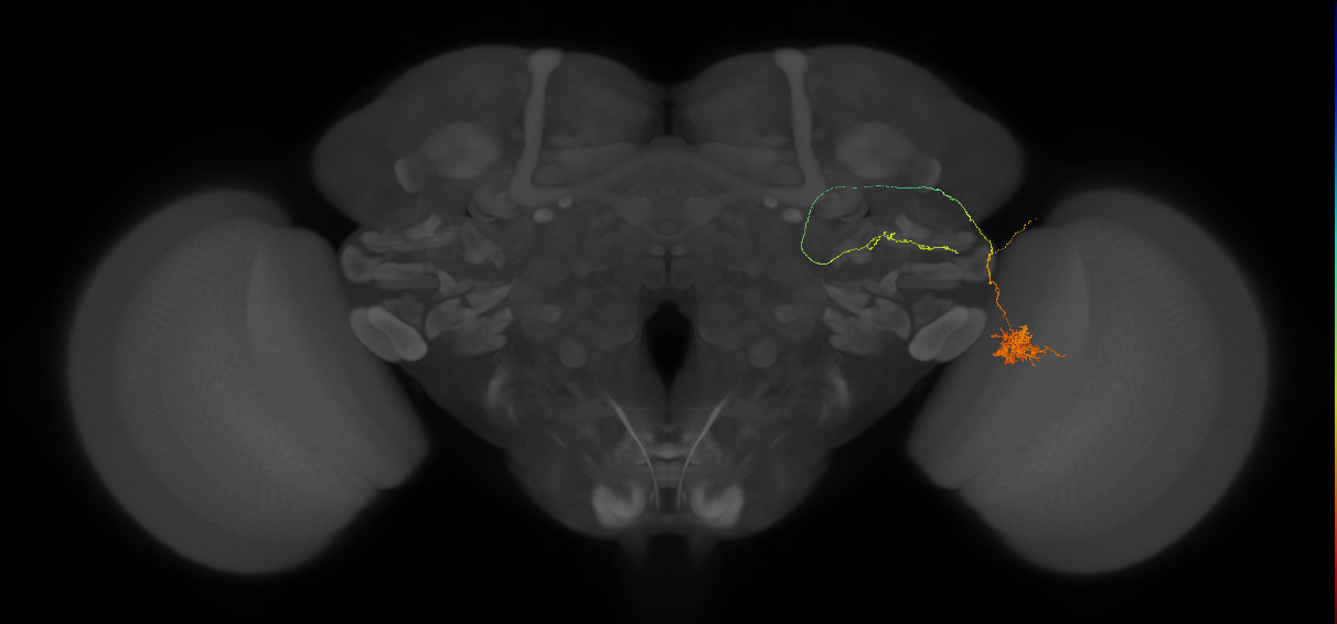
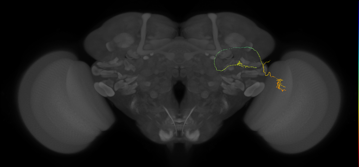
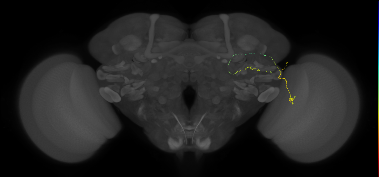
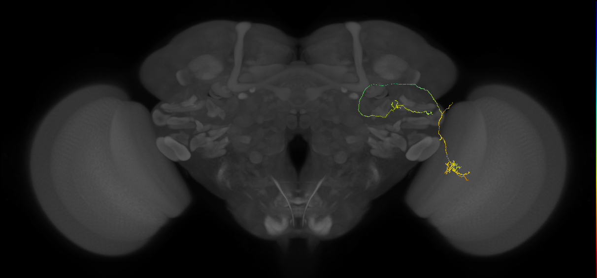
EM FAFB Morimoto et al 2020
ID: Morimoto2020
FAFB EM reconstructed neurons from Morimoto et al 2020
Open in VFB 3D Browser →Cross References
Imaging Technique
- transmission electron microscopy (TEM)
Downloads
Image files aligned to LC6#56 (FAFB:3322741):
Image files aligned to LC6#50 (FAFB:3301787):
Image files aligned to LC6#45 (FAFB:3299153):
Image files aligned to LC6#41 (FAFB:3297737):
Image files aligned to LC6#29 (FAFB:3288702):
Image files aligned to LC6#28 (FAFB:3286896):
Image files aligned to LC6#16 (FAFB:3267027):
Image files aligned to LC6#12 (FAFB:3162746):
Image files aligned to LC6G1#4 (FAFB:3155729):
Image files aligned to LC6#3 (FAFB:3117105):
References
29 - EM FAFB Otto et al 2020 [Otto2020]
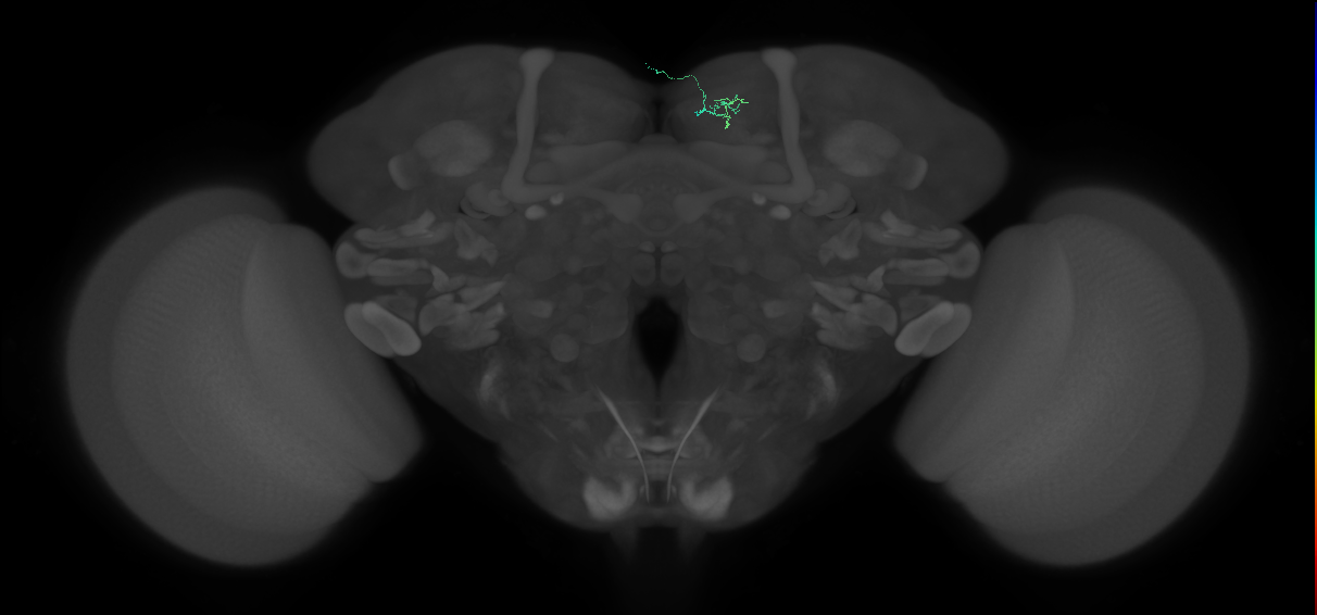
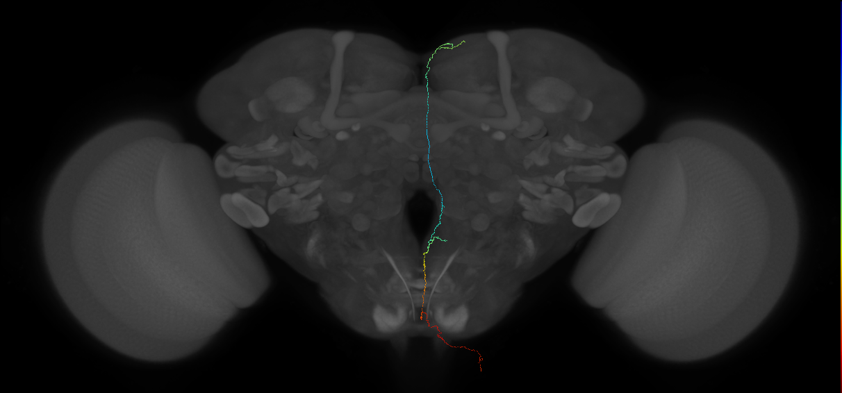
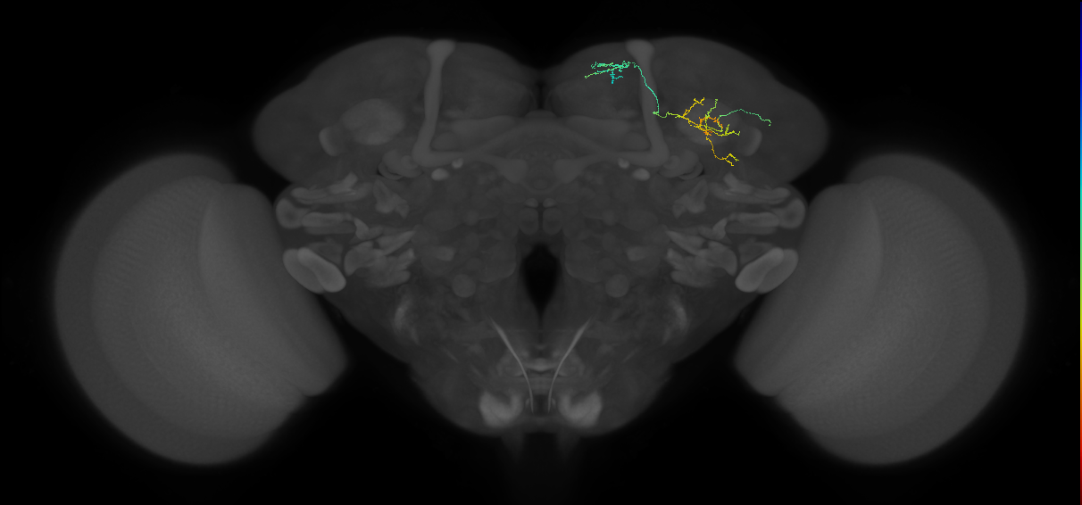
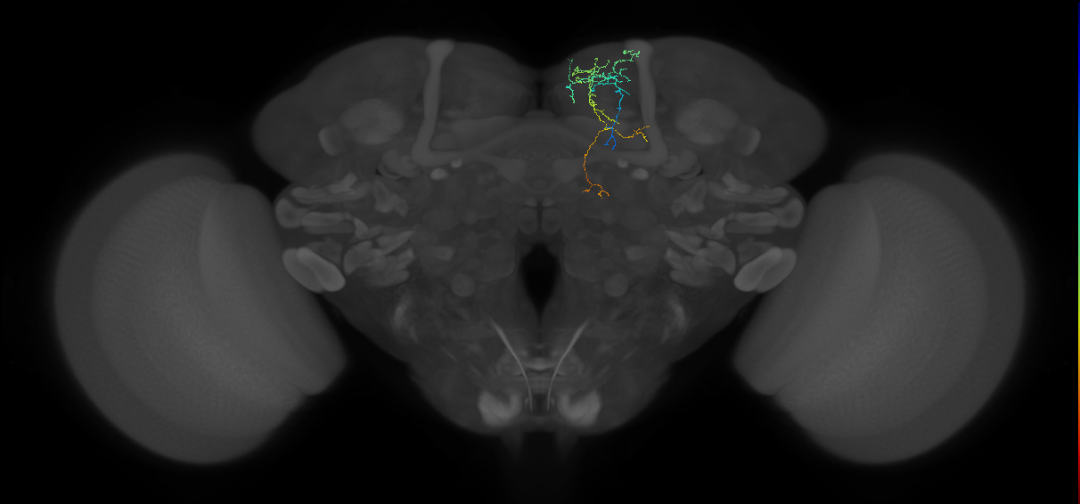
EM FAFB Otto et al 2020
ID: Otto2020
FAFB EM reconstructed neurons from Otto et al 2020
Open in VFB 3D Browser →Cross References
Imaging Technique
- transmission electron microscopy (TEM)
Downloads
Image files aligned to Protocerebral_DAN_input_c 3.8.0#8 (FAFB:8715837):
Image files aligned to Protocerebral_DAN_input_c 5.2.0#3 (FAFB:7532738):
Image files aligned to Protocerebral_DAN_input_c 10.12.0#2 (FAFB:7021238):
Image files aligned to Protocerebral_DAN_input_c 3.24.4 (FAFB:6036782):
Image files aligned to Protocerebral_DAN_input_c 12.10.0#5 (FAFB:5888228):
Image files aligned to Protocerebral_DAN_input_c 17.4.41#1 (FAFB:5073219):
Image files aligned to Protocerebral_DAN_input_c 9.4.1#3 (FAFB:4592543):
Image files aligned to Protocerebral_DAN_input_c 15.12.2 (FAFB:3183911):
Image files aligned to DAN_USN_c 11.9.3#1 (FAFB:1945705):
Image files aligned to Multiglomerular mlALT vPN#R1 (FAFB:3003322):
Images in dataset EM FAFB Otto et al 2020 (807 total)
| Thumbnail | Name | Tags |
|---|---|---|
 | [Protocerebral_DAN_input_c 17.4.41#11 (FAFB:8422016)](/reports/VFB_001014la) | Nervous_system, Neuron, Adult |
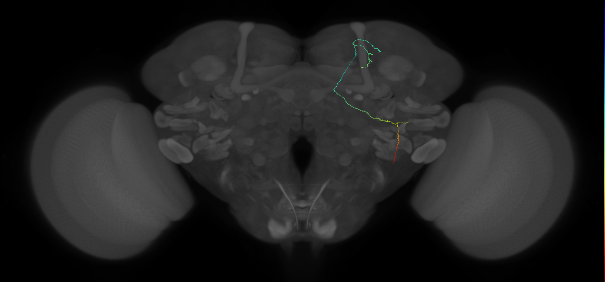
 | [Protocerebral_DAN_input_c 10.10.7#4 (FAFB:6834056)](/reports/VFB_001014ia) | Nervous_system, Neuron, Adult |
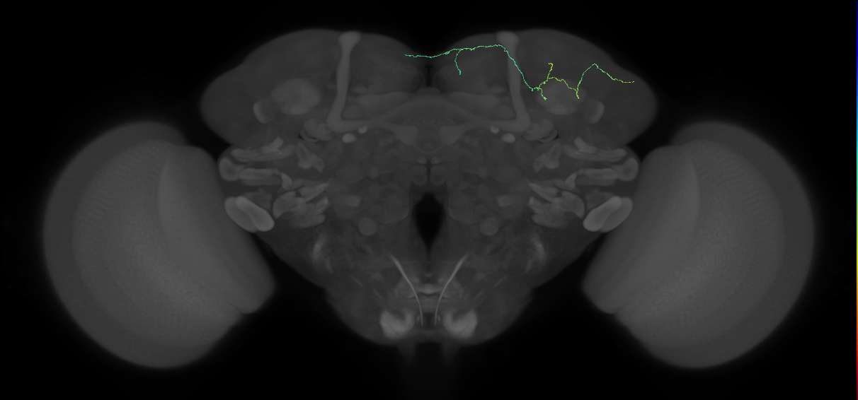
 | [SEZ_DAN_input_c 8.4.0 (FAFB:6423281)](/reports/VFB_001014hq) | Nervous_system, Neuron, Adult |
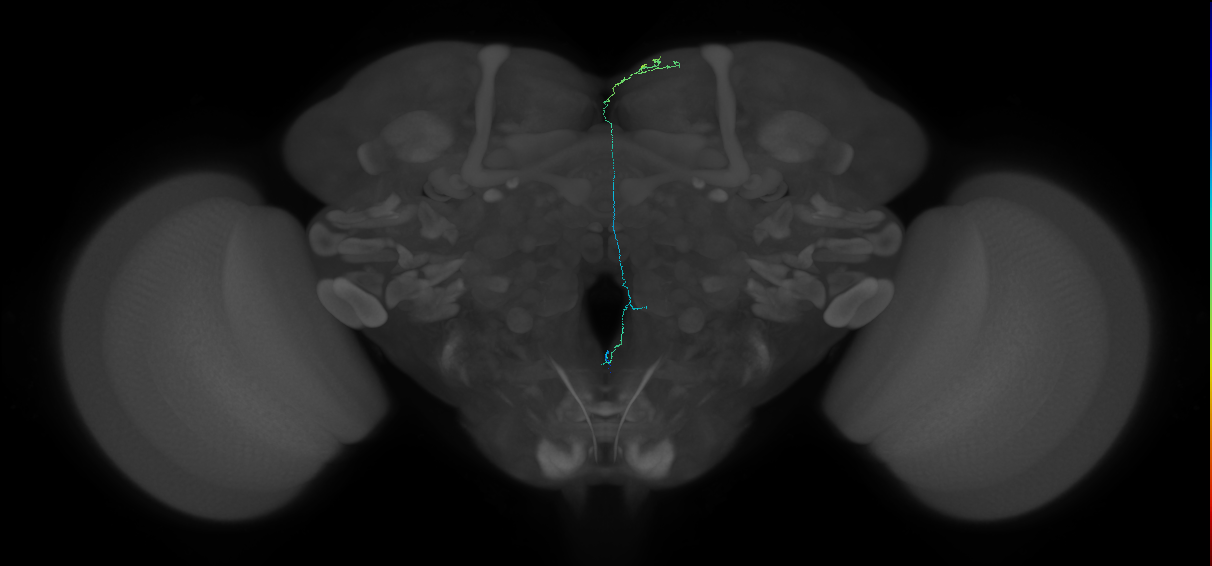 | [Protocerebral_DAN_input_c 5.3.0 (FAFB:6084281)](/reports/VFB_001014gl) | Nervous_system, Neuron, Adult |
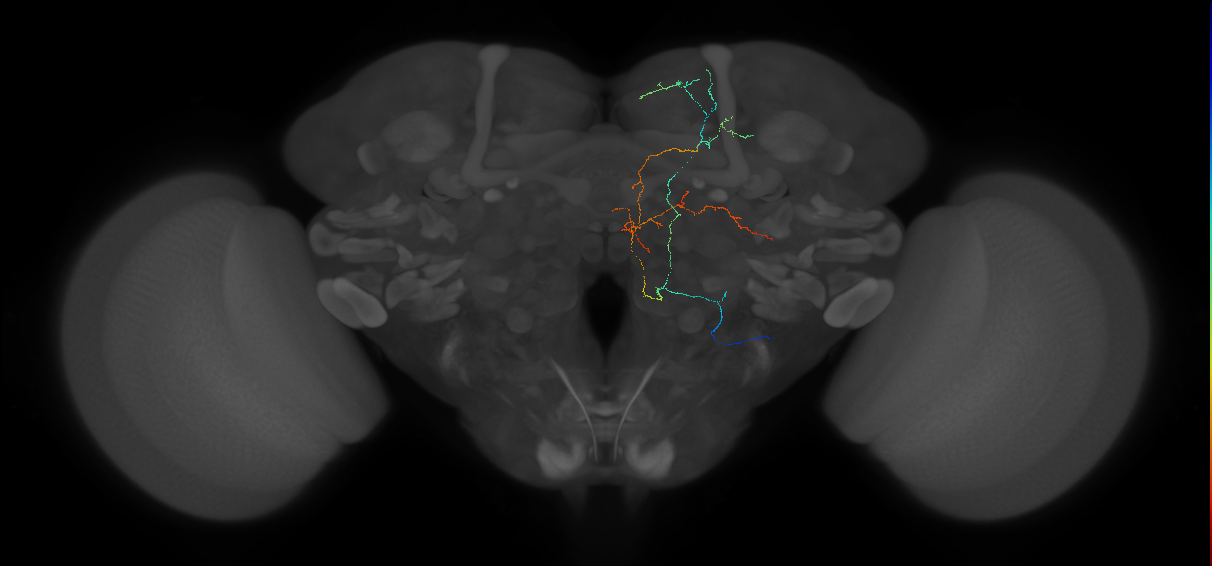 | [DAN_USN_c 1.2.0 (FAFB:5961542)](/reports/VFB_001014fs) | Nervous_system, Neuron, Adult |
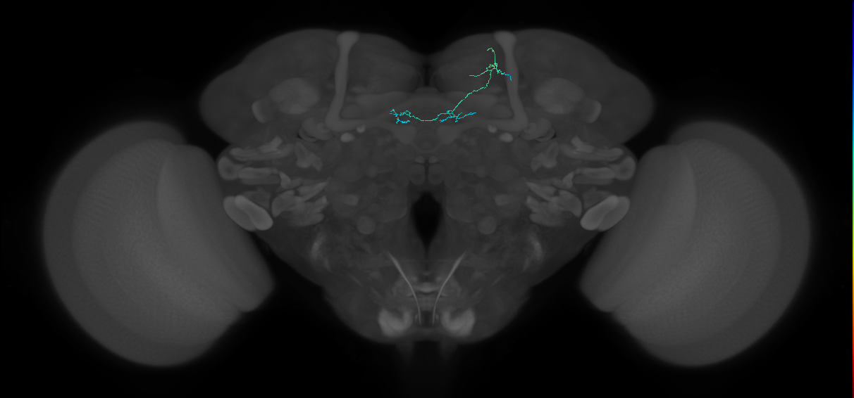 | [PAM-b'2a#2_R (FAFB:5886218)](/reports/VFB_001014f8) | secondary_neuron, Nervous_system, Adult, Dopaminergic |
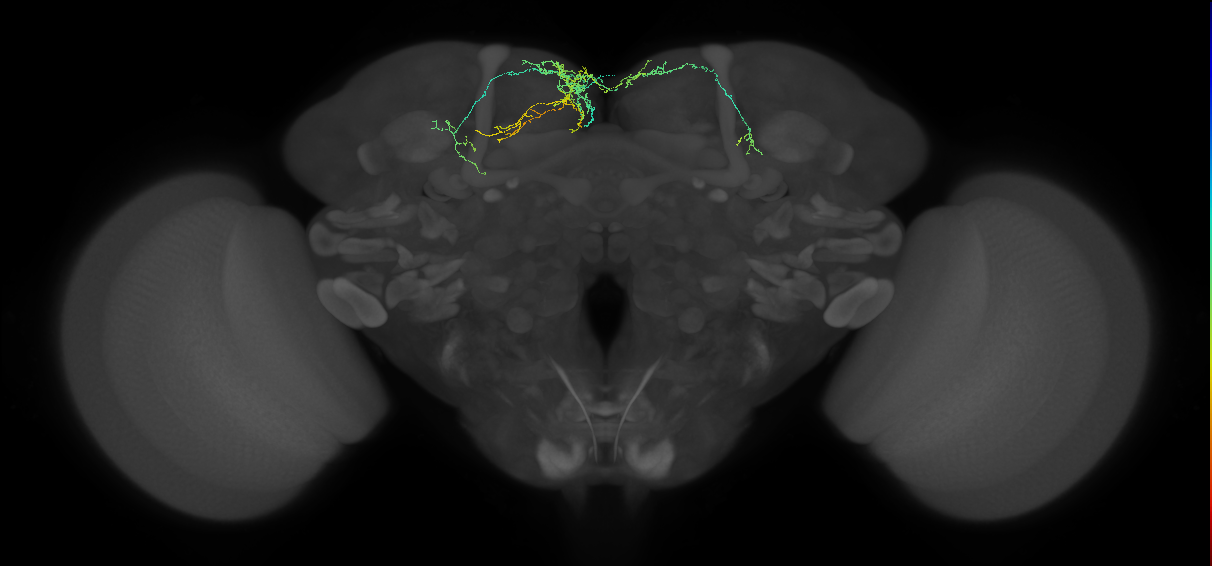 | [Protocerebral_DAN_input_c 3.10.0#1 (FAFB:3205031)](/reports/VFB_0010148z) | Nervous_system, Neuron, Adult |
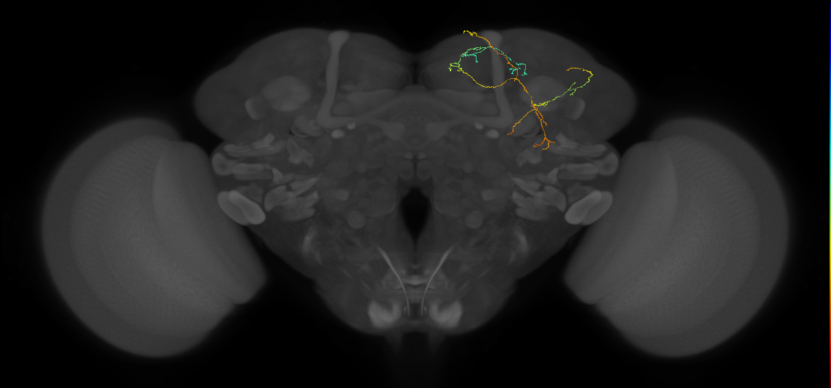 | [Protocerebral_DAN_input_c 12.8.0#4 (FAFB:3075536)](/reports/VFB_0010146q) | Nervous_system, Neuron, Adult |
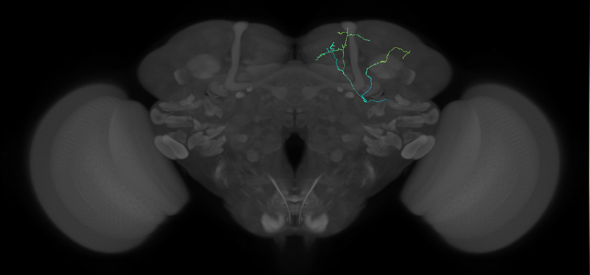 | [Protocerebral_DAN_input_c 15.11.2#1 (FAFB:2956363)](/reports/VFB_0010145u) | Nervous_system, Neuron, Adult |
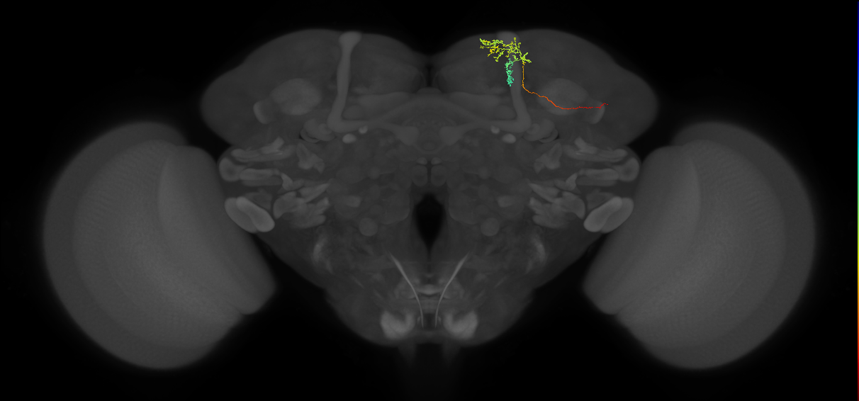 | [MBON-a2p3p#2_R (FAFB:1305588)](/reports/VFB_0010141f) | secondary_neuron, Adult, lineage_DL1_CP2, Cholinergic |
References
30 - EM FAFB Sayin et al 2019 [Sayin2019]


EM FAFB Sayin et al 2019
ID: Sayin2019
FAFB EM reconstructed neurons from Sayin et al 2019
Open in VFB 3D Browser →Cross References
Imaging Technique
- transmission electron microscopy (TEM)
Downloads
Image files aligned to MBON-y1pedc>a/B_R (FAFB:2333007):
Image files aligned to OA-VPM3 (FAFB:1329078):
Image files aligned to OA-VPM4 (FAFB:1191261):
References
31 - EM FAFB Shiu et al. 2022 [Shiu2022]
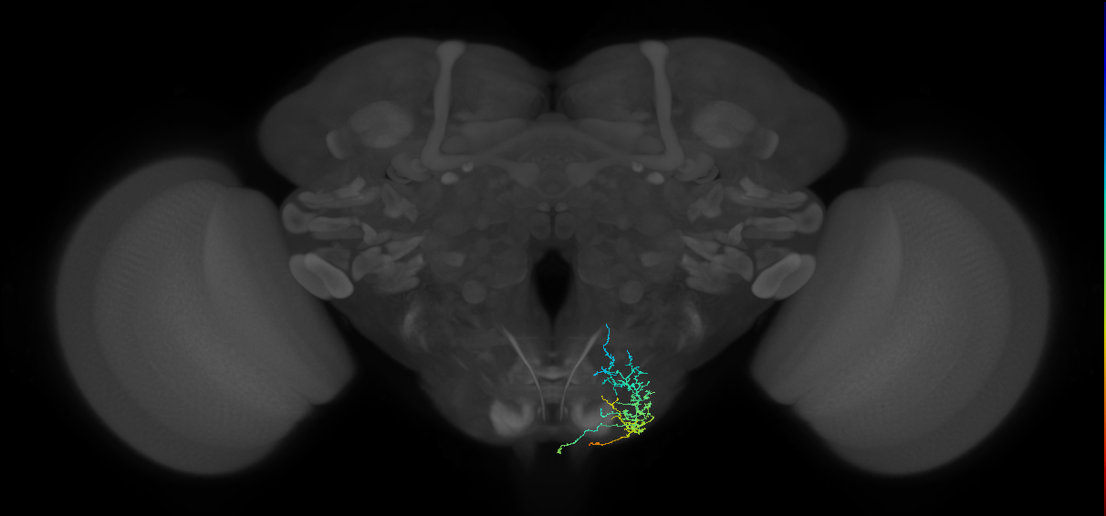
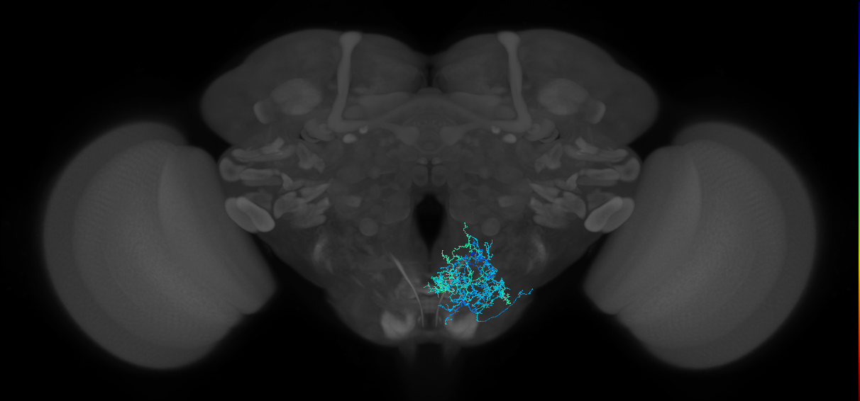
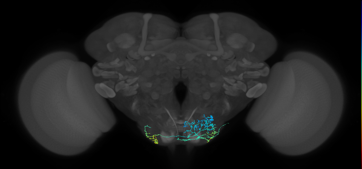
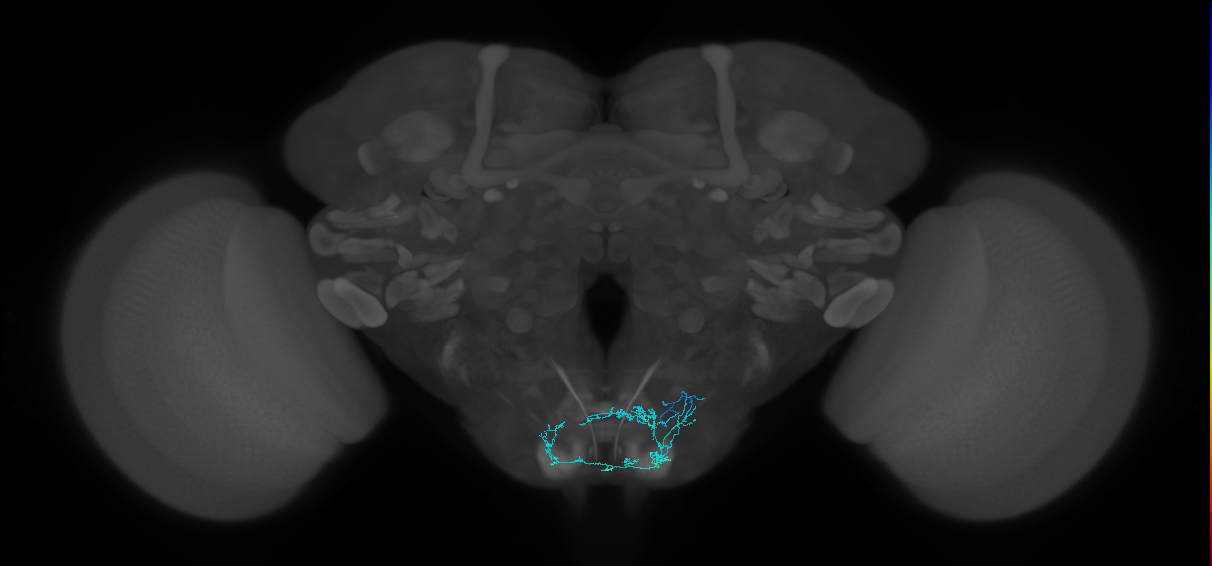
EM FAFB Shiu et al. 2022
ID: Shiu2022
FAFB EM reconstructed neurons from Shiu et al 2022
Open in VFB 3D Browser →Cross References
Imaging Technique
- transmission electron microscopy (TEM)
Downloads
Image files aligned to Bract#1 (FAFB:17024882):
Image files aligned to Scapula#1 (FAFB:16887116):
Image files aligned to Rounddown#1 (FAFB:16886973):
Image files aligned to G2N-1#1 (FAFB:15079937):
Image files aligned to Usnea#1 (FAFB:14890522):
Image files aligned to Clavicle#1 (FAFB:10150139):
Image files aligned to FMIN#1 (FAFB:8952676):
Image files aligned to Quasimodo#1 (FAFB:8275570):
Image files aligned to Buster#1 (FAFB:7899207):
Image files aligned to Zorro#L1 (FAFB:7574284):
References
32 - EM FAFB Taisz and Galili et al., 2022 [TaiszGalili2022]
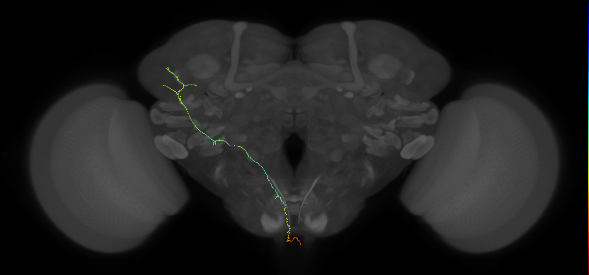


EM FAFB Taisz and Galili et al., 2022
ID: TaiszGalili2022
FAFB EM reconstructed neurons from Taisz and Galili et al., 2022
Open in VFB 3D Browser →Cross References
Imaging Technique
- transmission electron microscopy (TEM)
Downloads
Image files aligned to G2N-aSLP_L#1 (FAFB:11463951):
Image files aligned to aSP-g3_L#1 (FAFB:7856299):
Image files aligned to pGRN_L (FAFB:7108314):
Image files aligned to AD1g1_R (FAFB:1316642):
Image files aligned to G2N-SLP4_R (FAFB:1102458):
Image files aligned to SEZ_DAN_input_c 7.4.0#1 (FAFB:1059970):
Image files aligned to pC1a (FAFB:3807212):
Image files aligned to Uniglomerular mALT DA1 lPN#L4 (FAFB:2379517):
Image files aligned to Uniglomerular mALT DA1 lPN#R7 (FAFB:57353):
Image files aligned to Multiglomerular mALT lvPN#R46 (FAFB:57179):
References
33 - EM FAFB Turner-Evans et al 2020 [Turner_Evans2020]

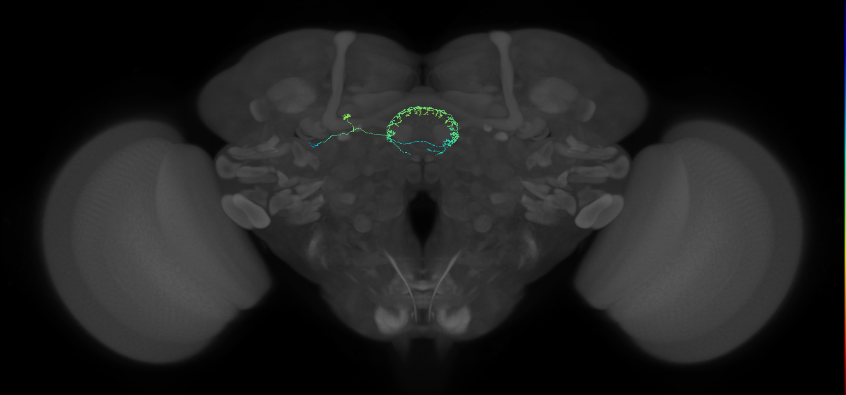
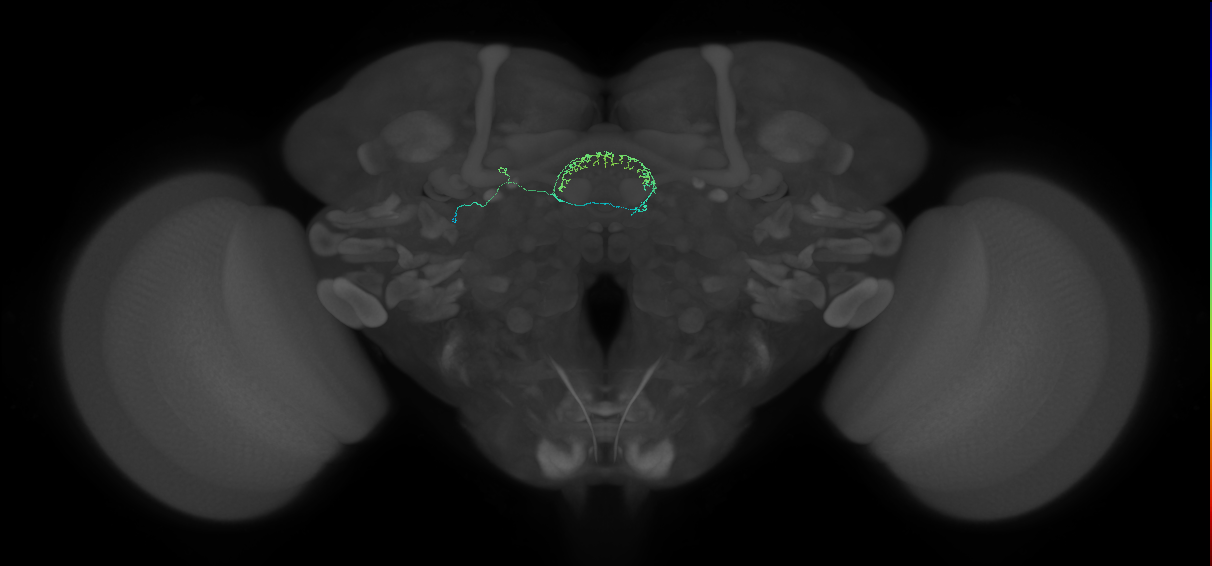
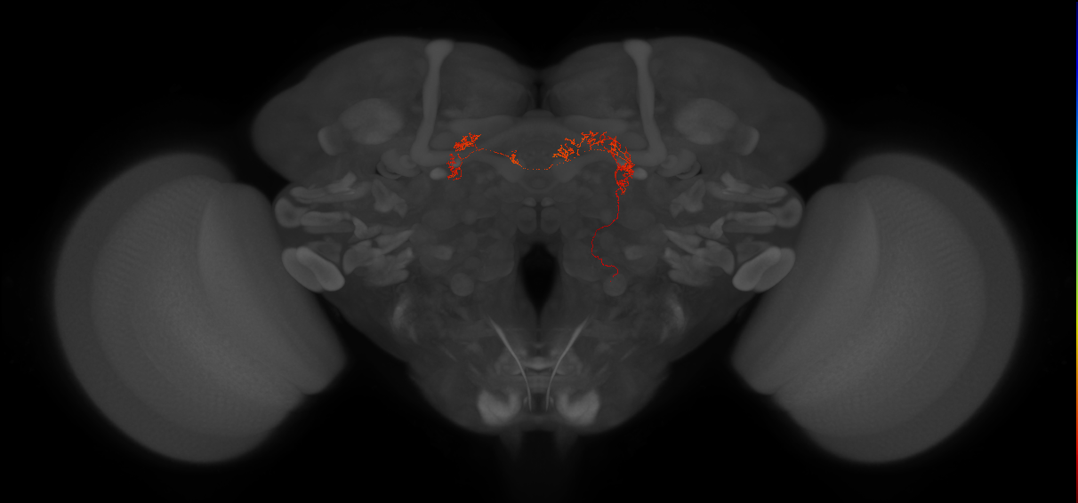
EM FAFB Turner-Evans et al 2020
ID: Turner_Evans2020
FAFB EM reconstructed neurons from Turner-Evans et al 2020
Open in VFB 3D Browser →Cross References
Imaging Technique
- transmission electron microscopy (TEM)
Downloads
Image files aligned to R4d#4 (FAFB:6813859):
Image files aligned to R4d#3 (FAFB:6417166):
Image files aligned to R4d#8 (FAFB:6393609):
Image files aligned to Delta7-7R2L#1 (FAFB:6254861):
Image files aligned to PEN1-6L#2 (FAFB:3423772):
Image files aligned to PEG-5L (FAFB:1032767):
Image files aligned to EPG-5R#2 (FAFB:861090):
Image files aligned to neuron_fragment#1 (FAFB:854144):
Image files aligned to PEN2-5R (FAFB:806288):
Image files aligned to PEN2-6R#1 (FAFB:659759):
References
34 - EM FAFB Wang et al 2020a [Wang2020a]


EM FAFB Wang et al 2020a
ID: Wang2020a
FAFB EM reconstructed neurons from Wang et al 2020a
Open in VFB 3D Browser →Cross References
Imaging Technique
- transmission electron microscopy (TEM)
Downloads
Image files aligned to oviIN (FAFB:13325634):
Image files aligned to SAG#R (FAFB:5353953):
Image files aligned to oviDNa#1 (FAFB:5143346):
Image files aligned to SAG#L (FAFB:4358524):
Image files aligned to pC1c (FAFB:3794183):
Image files aligned to pC1b (FAFB:3781621):
Image files aligned to pC1e (FAFB:3770805):
Image files aligned to oviDNa#2 (FAFB:1875104):
Image files aligned to oviDNb (FAFB:1862762):
Image files aligned to oviEN (FAFB:1259226):
References
35 - EM FAFB Wang et al 2020b [Wang2020b]

EM FAFB Wang et al 2020b
ID: Wang2020b
FAFB EM reconstructed neurons from Wang et al 2020b
Open in VFB 3D Browser →Cross References
Imaging Technique
- transmission electron microscopy (TEM)
Downloads
Image files aligned to DNp13 (FAFB:5321581):
References
36 - EM FAFB Wang et al 2020c [Wang2020c]
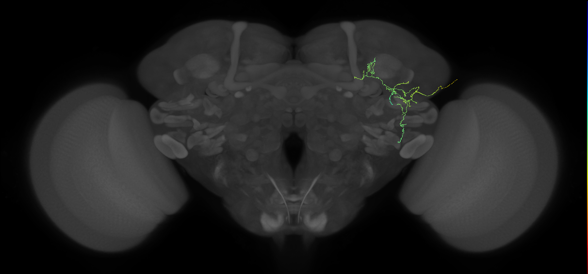
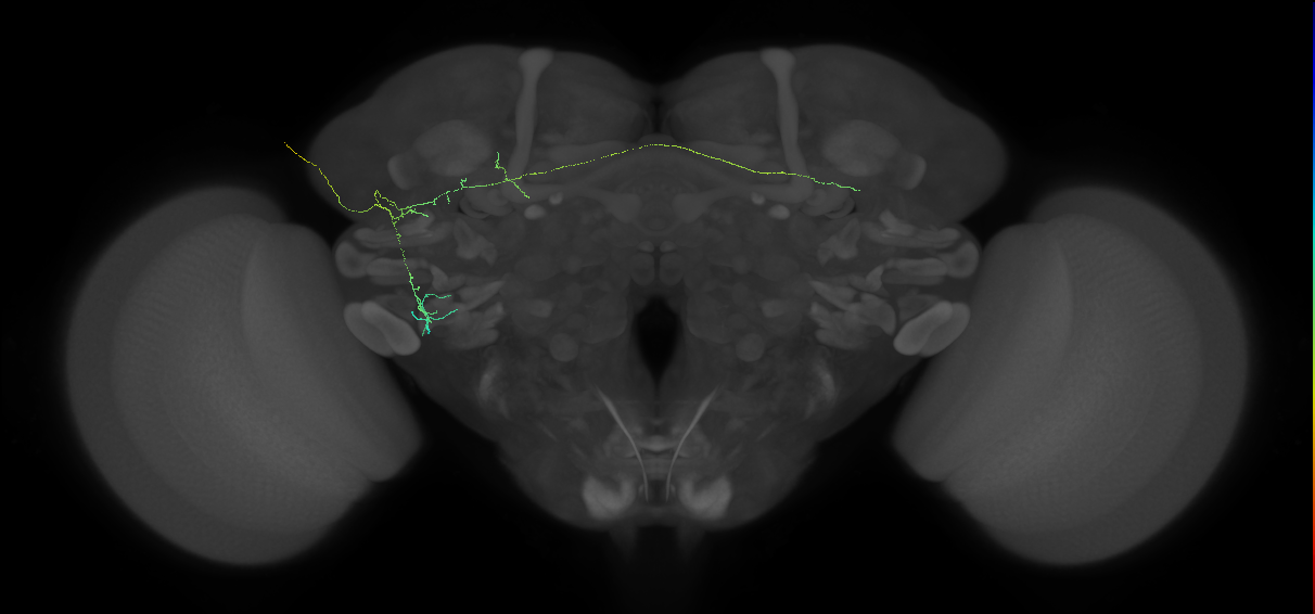
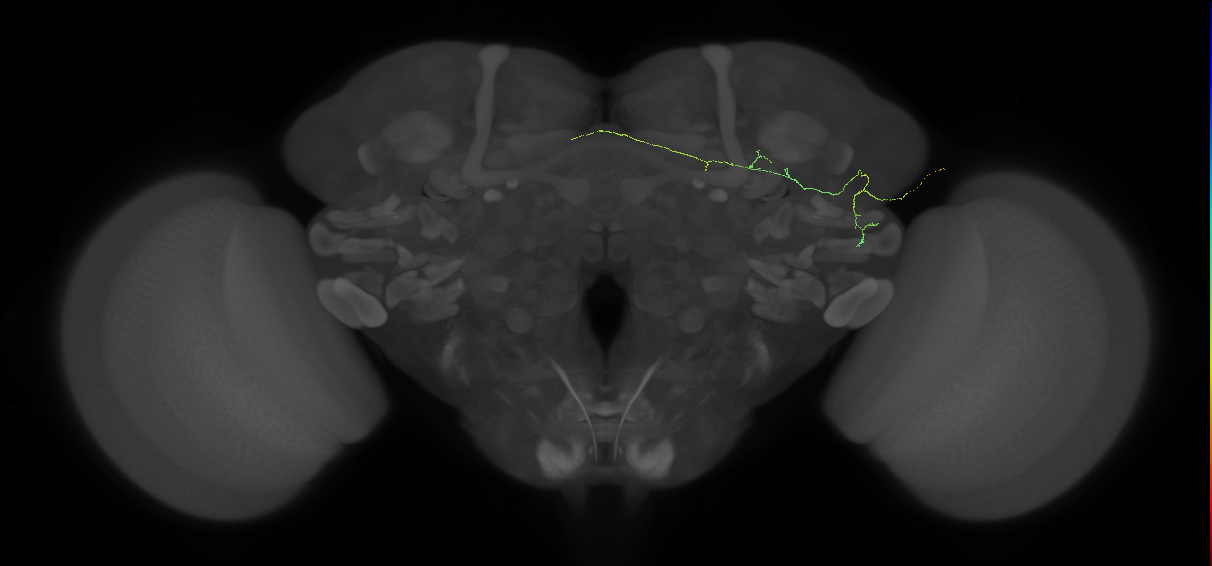
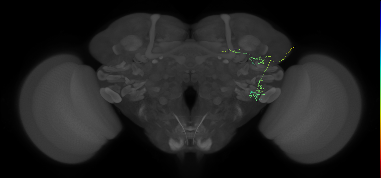
EM FAFB Wang et al 2020c
ID: Wang2020c
FAFB EM reconstructed neurons from Wang et al 2020c
Open in VFB 3D Browser →Cross References
Imaging Technique
- transmission electron microscopy (TEM)
Downloads
Image files aligned to vpoIN#7 (FAFB:7125205):
Image files aligned to vpoIN#11 (FAFB:6540243):
Image files aligned to vpoIN#14 (FAFB:6031288):
Image files aligned to vpoIN#8 (FAFB:5753206):
Image files aligned to vpoIN#9 (FAFB:5592796):
Image files aligned to SAG#L (FAFB:4358524):
Image files aligned to pC1d (FAFB:3979930):
Image files aligned to pC1c (FAFB:3794183):
Image files aligned to vpoEN#2 (FAFB:5480914):
Image files aligned to vpoIN#10 (FAFB:5123560):
References
37 - EM FAFB Zhao et al., 2023 [Zhao2023]
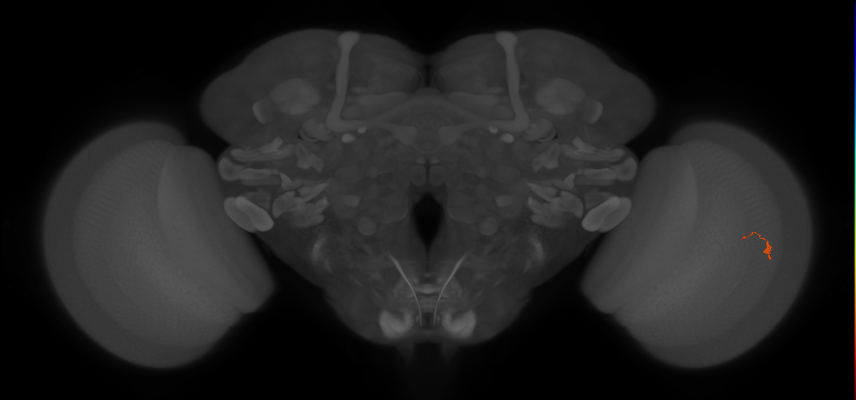
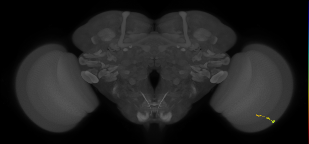
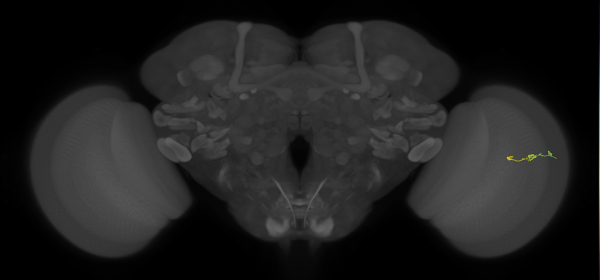
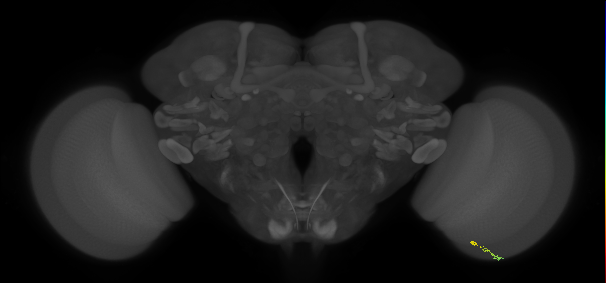
EM FAFB Zhao et al., 2023
ID: Zhao2023
FAFB EM reconstructed neurons from Zhao et al., 2023
Open in VFB 3D Browser →Cross References
Imaging Technique
- transmission electron microscopy (TEM)
Downloads
Image files aligned to T4c_R#21 (FAFB:17018569):
Image files aligned to Mi1_R#729 (FAFB:15421511):
Image files aligned to Mi1_R#400 (FAFB:14890488):
Image files aligned to Mi1_R#236 (FAFB:14788007):
Image files aligned to Mi1_R#160 (FAFB:13689085):
Image files aligned to T4b_R#149 (FAFB:11760844):
Image files aligned to T4c_R#10 (FAFB:11279787):
Image files aligned to Mi1_R#15 (FAFB:10148425):
Image files aligned to T4b_R#102 (FAFB:9996235):
Image files aligned to T4d_R#30 (FAFB:8453990):
References
38 - EM FAFB Zheng et al 2018 [Zheng2018]

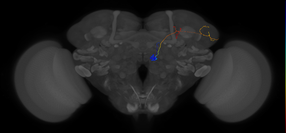


EM FAFB Zheng et al 2018
ID: Zheng2018
FAFB EM reconstructed neurons from Zheng et al 2018
Open in VFB 3D Browser →Cross References
Imaging Technique
- transmission electron microscopy (TEM)
Downloads
Image files aligned to Uniglomerular mALT VM5d adPN#R3 (FAFB:62434):
Image files aligned to Uniglomerular mALT VM5d adPN#R1 (FAFB:39139):
Image files aligned to Uniglomerular mALT DM4 adPN#R1 (FAFB:39682):
Image files aligned to Uniglomerular mALT DA4l adPN#R1 (FAFB:40749):
Image files aligned to Uniglomerular mALT DL3 lPN#R5 (FAFB:33903):
Image files aligned to Uniglomerular mALT D adPN#R2 (FAFB:35246):
Image files aligned to Uniglomerular mALT DC1 adPN#R1 (FAFB:771242):
Image files aligned to Uniglomerular mALT VL2p adPN#R1 (FAFB:22976):
Image files aligned to Uniglomerular mALT VC3l adPN#R1 (FAFB:46800):
Image files aligned to Uniglomerular mALT VM7v adPN#R1 (FAFB:24251):
References
39 - EM FAFB Zheng et al 2020 (published 2022) [Zheng2020]
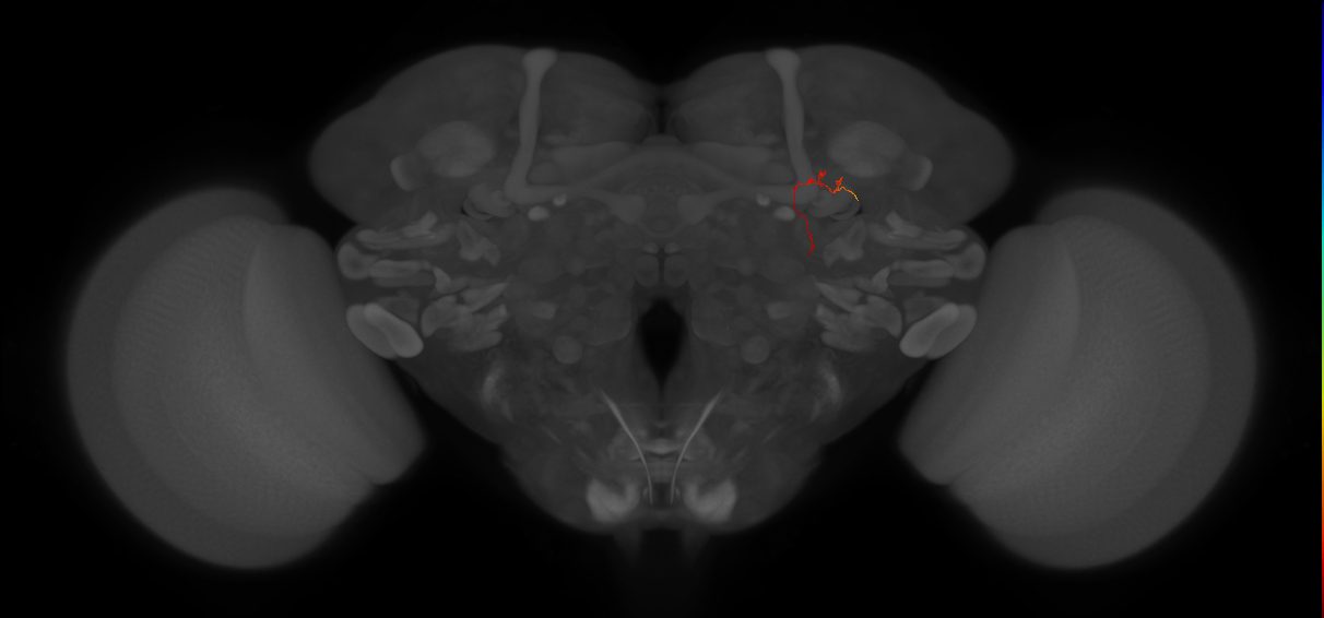
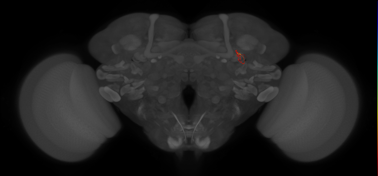
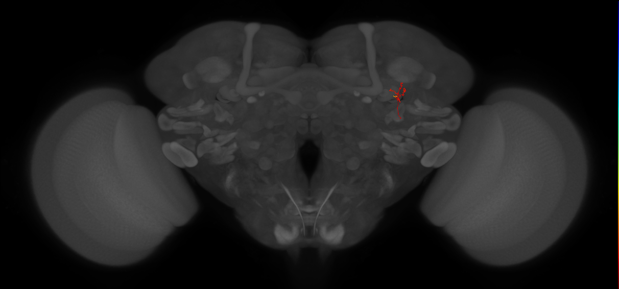
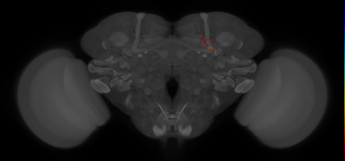
EM FAFB Zheng et al 2020 (published 2022)
ID: Zheng2020
FAFB EM reconstructed neurons from Zheng et al 2020 (published 2022)
Open in VFB 3D Browser →Cross References
Imaging Technique
- transmission electron microscopy (TEM)
Downloads
Image files aligned to KC#1466-y (FAFB:11006572):
Image files aligned to KC#1390-y (FAFB:10890313):
Image files aligned to KC#1378-ab (FAFB:10851177):
Image files aligned to KC#1124-a’b’ (FAFB:10120487):
Image files aligned to KC#1010-y (FAFB:9764382):
Image files aligned to KC#848-y (FAFB:9271871):
Image files aligned to KC#820-y (FAFB:9168453):
Image files aligned to KC#713-a’b’ (FAFB:8447556):
Image files aligned to KC#584-a’b’ (FAFB:2717438):
Image files aligned to KC#243-y (FAFB:16990):
References
40 - EM FANC Phelps et al 2020 [Maniates_Selvin2020]

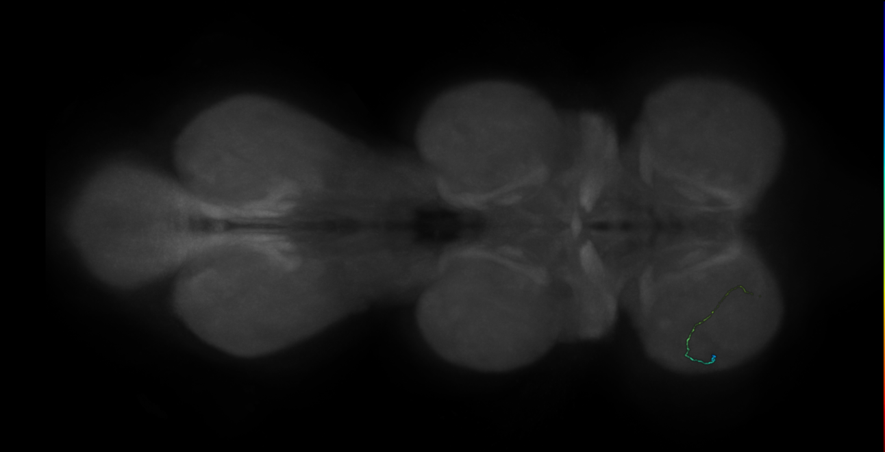
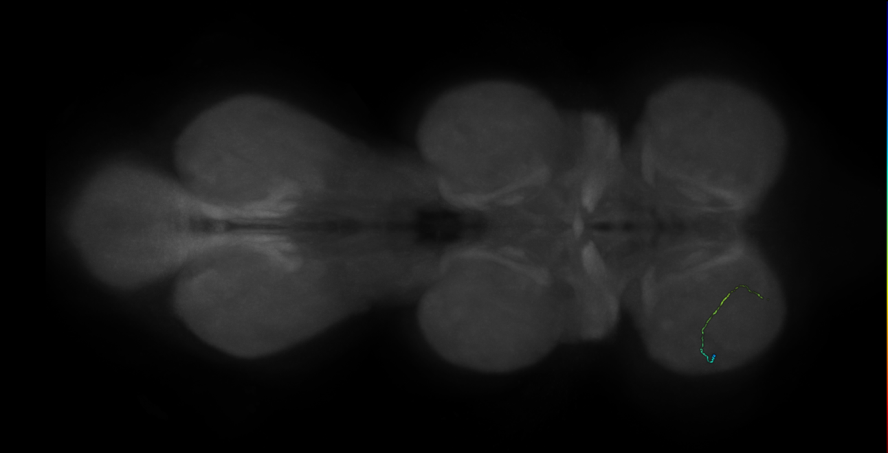
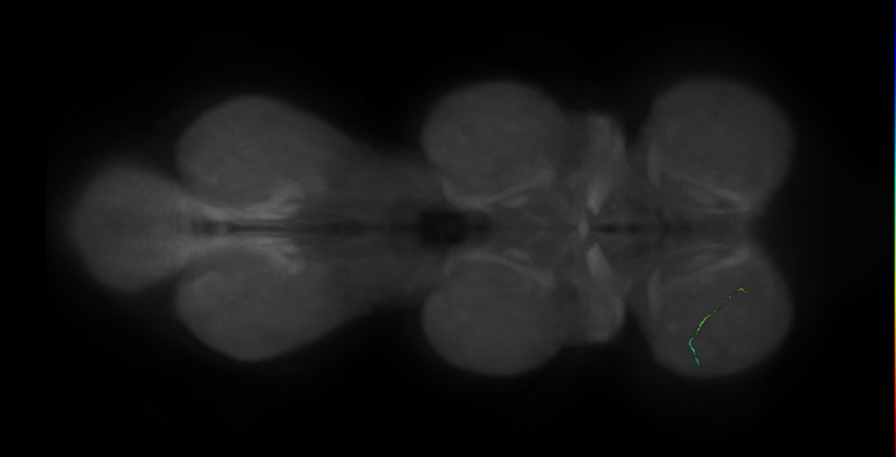
EM FANC Phelps et al 2020
ID: Maniates_Selvin2020
FANC EM reconstructed neurons from Maniates-Selvin et al 2020
Open in VFB 3D Browser →Imaging Technique
- transmission electron microscopy (TEM)
Downloads
Image files aligned to neuron 99449 - flipped (FANC:666363):
Image files aligned to neuron 9207 - radius 500 (FANC:597289):
Image files aligned to neuron 9191 - radius 500 (FANC:596827):
Image files aligned to neuron 10424 - pruned by vol 109 - radius 500 (FANC:594501):
Image files aligned to neuron 23722 (FANC:515106):
Image files aligned to neuron 73083 (FANC:499785):
Image files aligned to neuron 11608 (FANC:499720):
Image files aligned to neuron 9191 (FANC:498211):
Image files aligned to neuron 11043 (FANC:497053):
Image files aligned to neuron 581 (FANC:495816):
References
41 - EM L1 Andrade et al. 2019 [Andrade2019]
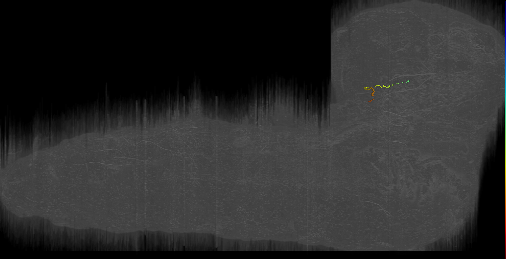
EM L1 Andrade et al. 2019
ID: Andrade2019
L1 EM reconstructed neurons from Andrade et al 2019
Open in VFB 3D Browser →Cross References
Imaging Technique
- transmission electron microscopy (TEM)
Downloads
Image files aligned to Close to BLVa s Left (L1EM:17728927):
Image files aligned to CP23 v s 5 Right (L1EM:17380305):
Image files aligned to BAlc post s 6 Left (L1EM:16461910):
Image files aligned to DALl1 s 4 Right (L1EM:14749848):
Image files aligned to CPd s 1 Right (L1EM:14547406):
Image files aligned to CM4 s loSM 2 Right (L1EM:14546829):
Image files aligned to BAmas12 ven s 4 Right (L1EM:13967121):
Image files aligned to CPf s 1 Right (L1EM:9816042):
Image files aligned to BLAd OL s 5 Left (L1EM:4663662):
Image files aligned to BLP34 b s 1 Right (L1EM:4127993):
References
42 - EM L1 Barnes et al., 2022 [Barnes2022]


EM L1 Barnes et al., 2022
ID: Barnes2022
L1 EM reconstructed neurons from Barnes et al., 2022
Open in VFB 3D Browser →Cross References
Imaging Technique
- transmission electron microscopy (TEM)
Downloads
Image files aligned to Drunken-1 a1r (L1EM:4096366):
Image files aligned to Ladder-f a2 (L1EM:7657358):
Image files aligned to chos_lch5-3_a1 (L1EM:8031834):
Image files aligned to chos_vchAB2_a1 (L1EM:7796832):
Image files aligned to chos_lch5-24-2_a1 (L1EM:6276557):
Image files aligned to Basin3_a1 (L1EM:3091943):
Image files aligned to Basin2_a1 (L1EM:3074106):
Image files aligned to chos_lch5-24-1_a1 (L1EM:162293):
Image files aligned to 45a PN (L1EM:8247451):
Image files aligned to _broad D1 (L1EM:7941652):
References
43 - EM L1 Carreira-Rosario, Arzan Zarin, Clark et al. 2018 [CarreiraRosario2018]


EM L1 Carreira-Rosario, Arzan Zarin, Clark et al. 2018
ID: CarreiraRosario2018
L1 EM reconstructed neurons from Carreira-Rosario, Arzan Zarin, Clark et al. 2018
Open in VFB 3D Browser →Cross References
Imaging Technique
- transmission electron microscopy (TEM)
Downloads
Image files aligned to A18b_a2r (L1EM:16002926):
Image files aligned to A27h_a7r (L1EM:15700463):
Image files aligned to MDNb_right (L1EM:13557064):
Image files aligned to ThDN right (L1EM:11541264):
Image files aligned to MDNa_left (L1EM:10728333):
Image files aligned to MDNb_left (L1EM:10728328):
Image files aligned to SEZ-DN1_left (L1EM:5308485):
Image files aligned to A18b_a1r (L1EM:4727546):
Image files aligned to A27l_a3l (L1EM:2111886):
Image files aligned to A27k_a1l (L1EM:4213417):
References
44 - EM L1 Eschbach et al 2020 [Eschbach2020]


EM L1 Eschbach et al 2020
ID: Eschbach2020
L1 EM reconstructed neurons from Eschbach et al 2020
Open in VFB 3D Browser →Cross References
Imaging Technique
- transmission electron microscopy (TEM)
Downloads
Image files aligned to FFN-5 (L1EM:3945454):
Image files aligned to FFN-15 (L1EM:16978882):
Image files aligned to FBN-5 (L1EM:15588620):
Image files aligned to FB2IN-1 (L1EM:12233237):
Image files aligned to CN-11 (L1EM:11909406):
Image files aligned to FBN-28 (L1EM:10612094):
Image files aligned to FB2IN-8 (L1EM:8540770):
Image files aligned to FFN-26 (L1EM:7058243):
Image files aligned to FFN-9 (L1EM:4665627):
Image files aligned to FFN-39 (L1EM:4116174):
References
45 - EM L1 Eschbach et al 2020b [Eschbach2020b]
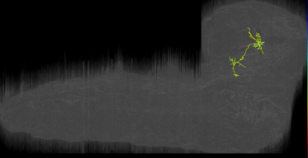
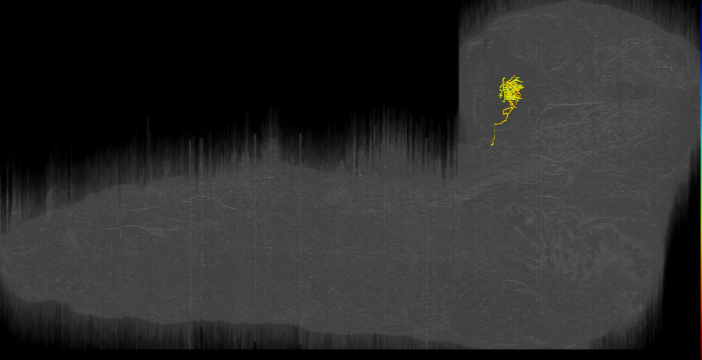
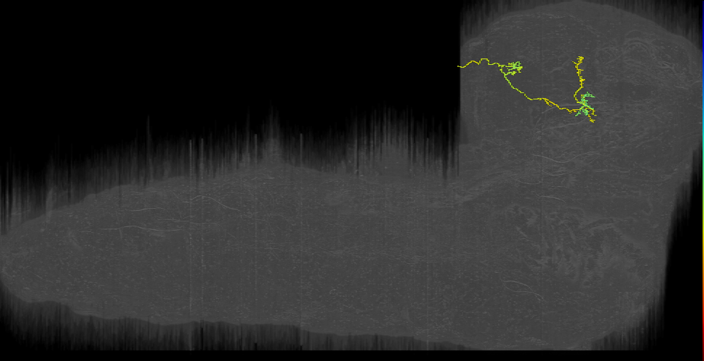
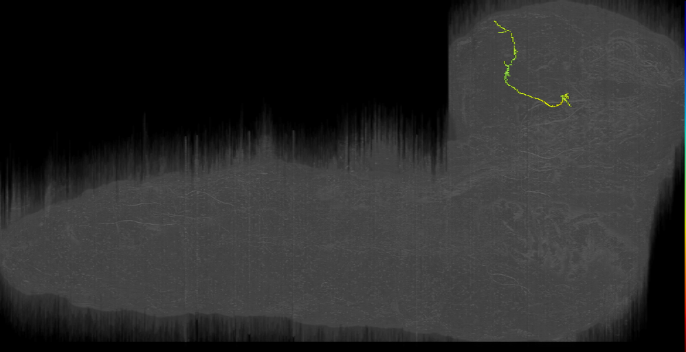
EM L1 Eschbach et al 2020b
ID: Eschbach2020b
L1 EM reconstructed neurons from Eschbach et al 2020b
Open in VFB 3D Browser →Cross References
Imaging Technique
- transmission electron microscopy (TEM)
Downloads
Image files aligned to CN-22 (L1EM:14071613):
Image files aligned to CN-49 (L1EM:4665186):
Image files aligned to KC (L1EM:17147410):
Image files aligned to KC (L1EM:16435406):
Image files aligned to KC (L1EM:11725127):
Image files aligned to KC (L1EM:8413663):
Image files aligned to KC (L1EM:4230061):
Image files aligned to KC (L1EM:3497901):
Image files aligned to KC (L1EM:29):
Image files aligned to CN-51 (L1EM:13803032):
References
46 - EM L1 Imambocus et al., 2022 [Imambocus2022]

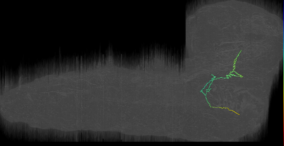

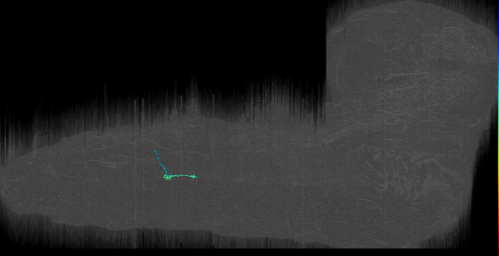
EM L1 Imambocus et al., 2022
ID: Imambocus2022
L1 EM reconstructed neurons from Imambocus et al., 2022
Open in VFB 3D Browser →Cross References
Imaging Technique
- transmission electron microscopy (TEM)
Downloads
Image files aligned to neuron 3918937 (L1EM:3918936):
Image files aligned to SEZ to brain left - gap todo (L1EM:16500055):
Image files aligned to AF6: Munin Output left (L1EM:3946364):
Image files aligned to vdaB_a4r (L1EM:9146908):
Image files aligned to v'td_vnc_a5l (L1EM:7012280):
Image files aligned to vdaB?_a6l (L1EM:1672957):
Image files aligned to vdaB_a3r (L1EM:1278501):
Image files aligned to vdaD class III md_a1r or es? (L1EM:2777891):
Image files aligned to v’td1_a1r (L1EM:10868477):
Image files aligned to MB2IN-202 (L1EM:4595609):
References
47 - EM L1 Jovanic et al. 2019 [Jovanic2019]


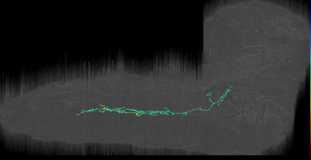
EM L1 Jovanic et al. 2019
ID: Jovanic2019
L1 EM reconstructed neurons from Jovanic et al., 2019
Open in VFB 3D Browser →Cross References
Imaging Technique
- transmission electron microscopy (TEM)
Downloads
Image files aligned to Thoracic Ladder-1 t2 (L1EM:20153309):
Image files aligned to A08h2_a2r (Jupiter right, not NB3-3) (L1EM:4228864):
Image files aligned to ddaA a1r class III md (L1EM:16312925):
Image files aligned to A09e_a4 (L1EM:183502):
Image files aligned to chos_vchAB2_a1 (L1EM:7796832):
Image files aligned to chos_lch5-1_a1 (L1EM:7764785):
Image files aligned to chos_lch5-5_a1 (L1EM:7763691):
Image files aligned to chos_v’ch_a1 (L1EM:7628068):
Image files aligned to chos_lch5-3_a1 (L1EM:7686464):
Image files aligned to chos_lch5-24-1_a1 (L1EM:7577773):
References
48 - EM L1 Mark et al. 2019 [Mark2019]


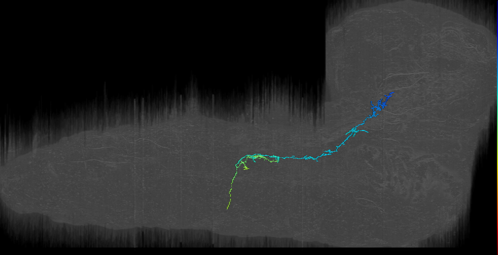

EM L1 Mark et al. 2019
ID: Mark2019
L1 EM reconstructed neurons from Mark et al. 2019
Open in VFB 3D Browser →Cross References
- EM L1 Mark et al. 2019 on VFB CATMAID L1 CNS (20726939)
Imaging Technique
- transmission electron microscopy (TEM)
Downloads
Image files aligned to A02e_a1l (L1EM:7992966):
Image files aligned to a03d/e_a1l (L1EM:6570401):
Image files aligned to NB5-2 lineage 6 (HMD X?)_a1l (L1EM:14522404):
Image files aligned to A01xxxxx_a1l (L1EM:8840084):
Image files aligned to A08j2_a1l (L1EM:8057753):
Image files aligned to A03xyz_a1l (L1EM:4039711):
Image files aligned to A08e1_a1l (L1EM:7876385):
Image files aligned to Griddle-2 a1l (L1EM:10804041):
Image files aligned to A08x_a1l (L1EM:5343578):
Image files aligned to A05q_a1l (L1EM:4740178):
References
49 - EM L1 Miroschnikow et al. 2018 [Miroschnikow2018]


EM L1 Miroschnikow et al. 2018
ID: Miroschnikow2018
L1 EM reconstructed neurons from Miroschnikow et al., 2018
Open in VFB 3D Browser →Cross References
Imaging Technique
- transmission electron microscopy (TEM)
Downloads
Image files aligned to SE0 L1 (L1EM:15995012):
Image files aligned to AN-R-Sens-B1-ACa-17 (L1EM:15548107):
Image files aligned to CN-12 (L1EM:14313750):
Image files aligned to AN-R-Sens-B3-ACp-09 (gustatory pheromone neuron) (L1EM:9853425):
Image files aligned to MB2IN-202 (L1EM:19120853):
Image files aligned to AN-R-Sens-B1-AVa-20 (L1EM:16442344):
Image files aligned to MB2ON-152 (L1EM:11868260):
Image files aligned to DH44 1 left (L1EM:10418394):
Image files aligned to Pre PMN right (L1EM:3436420):
Image files aligned to Multiglomerular PN 3 (L1EM:2612527):
References
50 - EM L1 Tastekin et al 2018 [Tastekin2018]
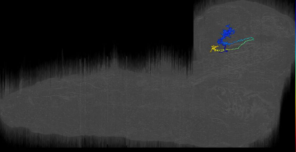
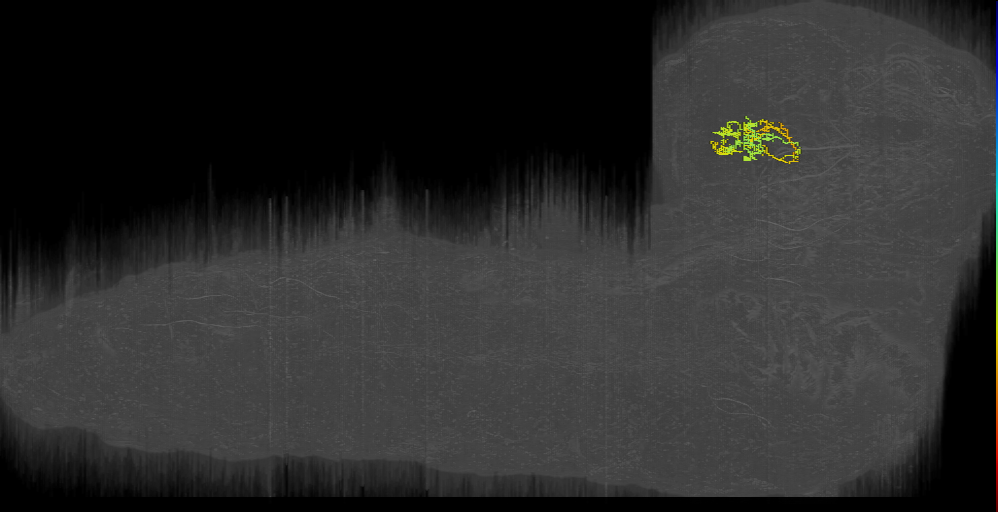


EM L1 Tastekin et al 2018
ID: Tastekin2018
L1 EM reconstructed neurons from Tastekin et al 2018
Open in VFB 3D Browser →Cross References
Imaging Technique
- transmission electron microscopy (TEM)
Downloads
Image files aligned to LHN-4 (L1EM:17806341):
Image files aligned to CN-27 (L1EM:17361757):
Image files aligned to CN-29 (L1EM:17038587):
Image files aligned to CN-27 (L1EM:12813847):
Image files aligned to LH-LN2_right (L1EM:9753418):
Image files aligned to CN-29 (L1EM:8065771):
Image files aligned to PDM-DN_left (L1EM:6446394):
Image files aligned to LH-LN2_left (L1EM:5769582):
Image files aligned to PDM-DN_right (L1EM:5462159):
Image files aligned to LHN-4 (L1EM:4680995):
References
51 - EM L1 Winding, Pedigo et al., 2023 [WindingPedigo2023]


EM L1 Winding, Pedigo et al., 2023
ID: WindingPedigo2023
L1 EM reconstructed neurons from Winding, Pedigo et al., 2023
Open in VFB 3D Browser →Cross References
Imaging Technique
- transmission electron microscopy (TEM)
Downloads
Image files aligned to lasso-top left (L1EM:17728723):
Image files aligned to Basal Tel-like 4 left (L1EM:16472086):
Image files aligned to MB2ON-260 (L1EM:12424940):
Image files aligned to BLAl_I-X 3 left (L1EM:12279941):
Image files aligned to AN-L-Sens-B1-AVa-32 (L1EM:1427012):
Image files aligned to ITP candidate 4 right (L1EM:7530253):
Image files aligned to CA-LP-2_left1 (L1EM:6978585):
Image files aligned to ITP candidate 1 left (L1EM:3925724):
Image files aligned to Rh6PR-4;right (L1EM:1856512):
Image files aligned to FB2IN-18 (L1EM:17176781):
References
52 - EM L1 Zarin, Mark et al. 2019 [Zarin2019]
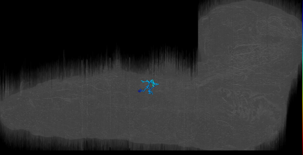


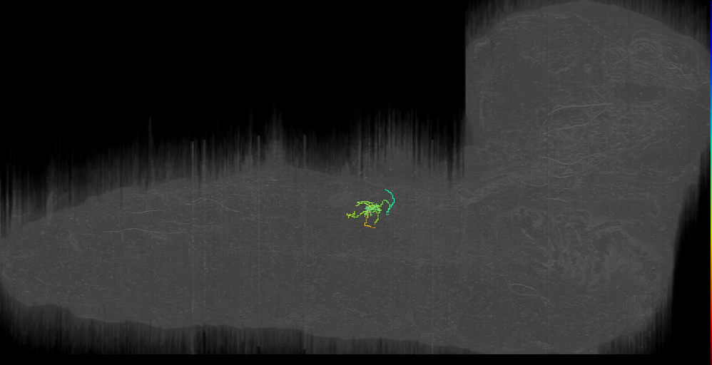
EM L1 Zarin, Mark et al. 2019
ID: Zarin2019
L1 EM reconstructed neurons from Zarin, Mark et al. 2019
Open in VFB 3D Browser →Cross References
Imaging Technique
- transmission electron microscopy (TEM)
Downloads
Image files aligned to MN8 (RF:SBM) LT3_a1r (L1EM:4881629):
Image files aligned to A27j_a1l (L1EM:17904627):
Image files aligned to A01c2_a1r (ipsi_dendrite) (L1EM:16226512):
Image files aligned to MN14 RP1_a1r (L1EM:14206438):
Image files aligned to MN14 RP1_a1l (L1EM:14045016):
Image files aligned to A01j_a1r (L1EM:4789092):
Image files aligned to MN26 (SN MN VA1)_a1r (L1EM:4397287):
Image files aligned to A08e1_a1l (L1EM:7876385):
Image files aligned to A05k_a1r (L1EM:10053304):
Image files aligned to A02m_a1r Pseudolooper-3 (L1EM:3546334):
References
53 - Eve+ neurons, sensorimotor circuit - EM (Heckscher2015) [Heckscher2015]
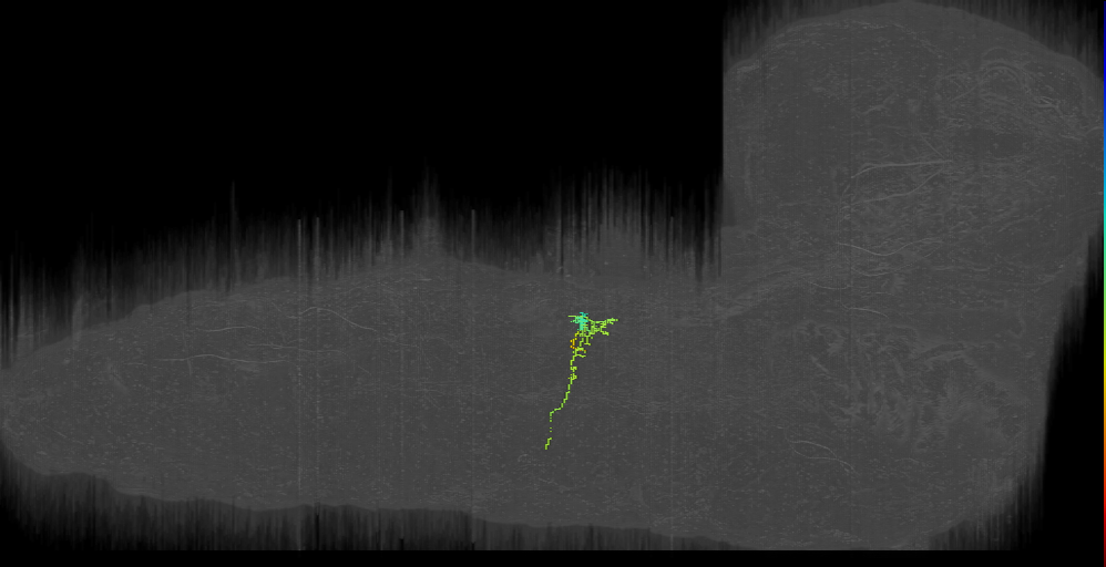
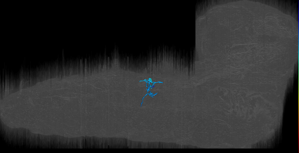


Eve+ neurons, sensorimotor circuit - EM (Heckscher2015)
ID: Heckscher2015
Cross References
Imaging Technique
- transmission electron microscopy (TEM)
Downloads
Image files aligned to MN15/16/17 VO4–6 (NB7-1? )_a1l (L1EM:19139253):
Image files aligned to MN22/23 (RF:LT2/LT3) LT1_a1r (L1EM:4102728):
Image files aligned to MNISNb/d-Is RP5_a1l (L1EM:14044614):
Image files aligned to MN12-III (ISN VL1 V-MN NB5-2)_a1l (L1EM:10678017):
Image files aligned to MN23/24 (RF: LT3/LT4) LT4_a1r (L1EM:4121534):
Image files aligned to neuron 11321645 frag run into a1 neuron (L1EM:11321644):
Image files aligned to MN1 aCC_a1r (L1EM:4488976):
Image files aligned to MN22/23 (RF:LT2/LT3) LT1_a1l (L1EM:4042315):
Image files aligned to A08e3_a2l (L1EM:10413917):
Image files aligned to A08e3_a1l (L1EM:8421329):
References
54 - FlyCircuit 1.0 - single neurons (Chiang2010) [Chiang2010]


Cross References
Imaging Technique
- confocal microscopy
Downloads
Image files aligned to Trh-F-200046:
Image files aligned to Cha-F-700048:
Image files aligned to TH-M-200080:
Image files aligned to Trh-M-500194:
Image files aligned to fru-M-900033:
Image files aligned to Trh-F-200046:
Image files aligned to Cha-F-700048:
Image files aligned to TH-M-200080:
Image files aligned to Trh-M-500194:
Image files aligned to fru-M-900033:
References
55 - FlyLight - Gen1 GAL4/LexA collection (exported 2019) [FlyLightGen1Set2019]
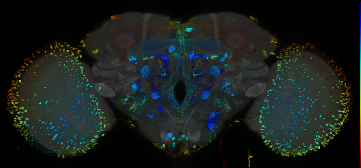


FlyLight - Gen1 GAL4/LexA collection (exported 2019)
ID: FlyLightGen1Set2019
Cross References
Imaging Technique
- confocal microscopy
Downloads
Image files aligned to R81G11:
Image files aligned to R12G04:
Image files aligned to R24G09L:
Image files aligned to R21G01L:
Image files aligned to R64E12L:
Image files aligned to R38G11L:
Image files aligned to R74E01:
Image files aligned to R27B12:
Image files aligned to R23E07:
Image files aligned to R18F04:
References
56 - FlyLight - GMR GAL4 collection (Jenett2012) [Jenett2012]


Cross References
Imaging Technique
- confocal microscopy
Downloads
Image files aligned to R16C12:
Image files aligned to R72G05:
Image files aligned to R93G11:
Image files aligned to R53H08:
Image files aligned to R18G02:
Image files aligned to R16C12:
Image files aligned to R72G05:
Image files aligned to R93G11:
Image files aligned to R53H08:
Image files aligned to R18G02:
References
57 - FlyLight split-GAL4 lines for Lateral Horn [FlyLight2019LateralHorn2019]


FlyLight split-GAL4 lines for Lateral Horn
ID: FlyLight2019LateralHorn2019
FlyLight split-GAL4 lines for Lateral Horn from Dolan et al., 2019
Open in VFB 3D Browser →Imaging Technique
- confocal microscopy
Downloads
Image files aligned to LH2088:
Image files aligned to LH2170:
Image files aligned to LH1899:
Image files aligned to LH1000:
Image files aligned to LH629:
Image files aligned to LH1827:
Image files aligned to LH412:
Image files aligned to LH2448:
Image files aligned to LH2094:
Image files aligned to LH1614:
References
58 - FlyLight split-GAL4 lines for Lateral Horn [LateralHorn2019]
FlyLight split-GAL4 lines for Lateral Horn
ID: LateralHorn2019
FlyLight split-GAL4 lines for Lateral Horn from Dolan et al., 2019
Open in VFB 3D Browser →References
59 - FlyWire connectome neurons [Dorkenwald2023]


FlyWire connectome neurons
ID: Dorkenwald2023
FlyWire Full Adult Female Brain (FAFB) connectome. Includes neuron skeletons, meshes and their synaptic connectivity
Open in VFB 3D Browser →Imaging Technique
- transmission electron microscopy (TEM)
Downloads
Image files aligned to C3:
Image files aligned to T5a:
Image files aligned to T4d:
Image files aligned to LPi4-3:
Image files aligned to T4b:
Image files aligned to T4d:
Image files aligned to KCa’b’-ap1:
Image files aligned to LoVP109:
Image files aligned to TuBu09:
Image files aligned to FB4:
References
60 - GAL4 lines from McKellar et al., 2020 [McKellar2020]


GAL4 lines from McKellar et al., 2020
ID: McKellar2020
GAL4 lines labelling proboscis muscle motor neurons from McKellar et al 2020
Open in VFB 3D Browser →Imaging Technique
- confocal microscopy
Downloads
Image files aligned to McKellar_SEZ_neurons_segmented_FDG:
Image files aligned to McKellar_Hugin:
Image files aligned to McKellar_Gr66a:
Image files aligned to McKellar_Gr64f:
References
61 - GAL4 Split expression patterns from Dolan et al. 2019 [Dolan2019]
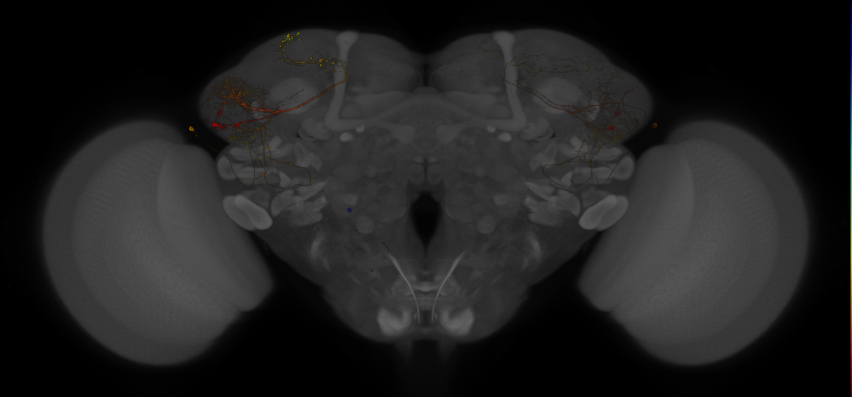

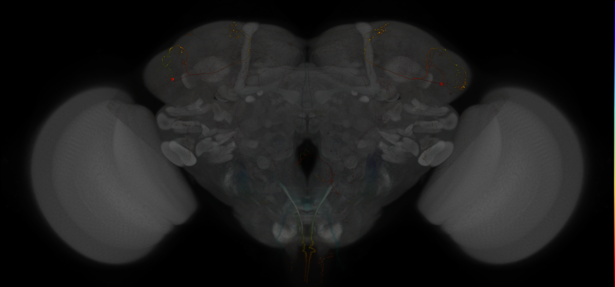
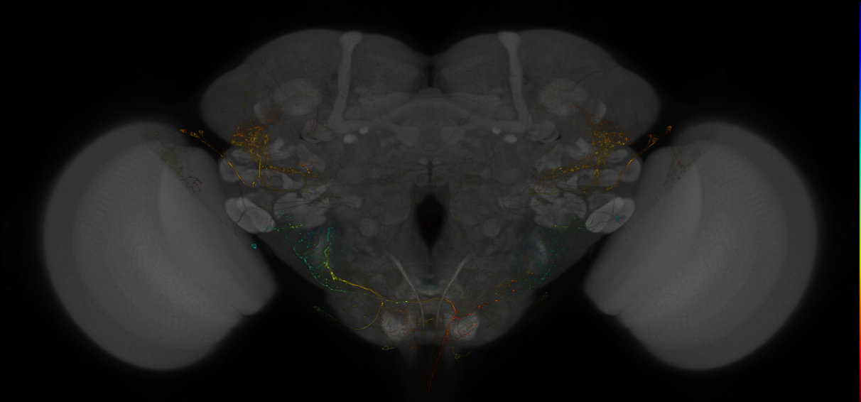
GAL4 Split expression patterns from Dolan et al. 2019
ID: Dolan2019
GAL4 Split expression patterns from Dolan et al., 2019
Open in VFB 3D Browser →Imaging Technique
- confocal microscopy
Downloads
Image files aligned to JRC_LH2456_Brain_20130201_19_E4_20x:
Image files aligned to LH18:
Image files aligned to LH1827:
Image files aligned to LH279:
Image files aligned to LH1354:
Image files aligned to LH1478:
Image files aligned to LH206:
Image files aligned to LH180:
Image files aligned to LH2446:
Image files aligned to LH2451:
References
62 - Grooming neurons and drivers (Hampel 2015) [FlyLight2019Hampel2015]
Grooming neurons and drivers (Hampel 2015)
ID: FlyLight2019Hampel2015
Images of grooming neurons and split GAL4 line drivers.
Open in VFB 3D Browser →References
63 - Grooming neurons and drivers (Hampel 2015) [Hampel2015]
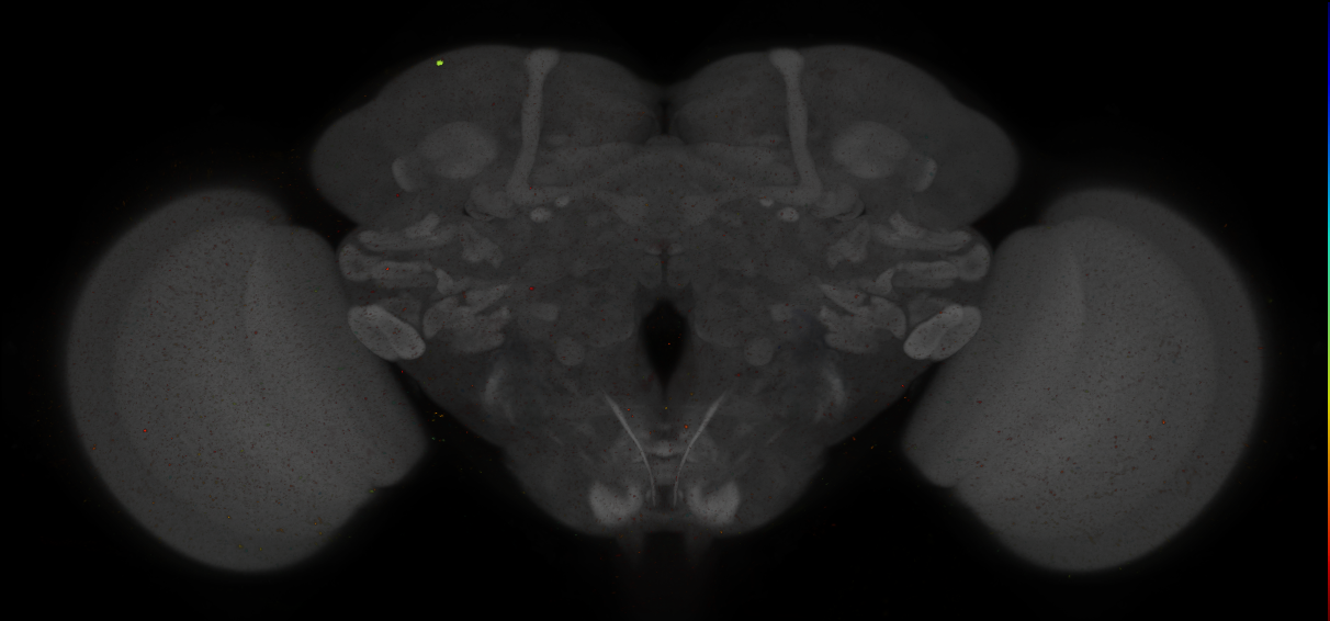
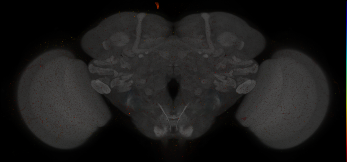


Grooming neurons and drivers (Hampel 2015)
ID: Hampel2015
Images of grooming neurons and split GAL4 line drivers.
Open in VFB 3D Browser →Imaging Technique
- confocal microscopy
Downloads
Image files aligned to spGAL4 control:
Image files aligned to spGAL4 control:
Image files aligned to aBN1-spGAL4-1 adult brain - Hampel2015:
Image files aligned to aDN1-spGAL4-1 adult brain - Hampel2015:
Image files aligned to aBN1-spGAL4-1 adult brain - Hampel2015:
Image files aligned to aDN1-spGAL4-1 adult brain - Hampel2015:
Image files aligned to spGAL4 control:
Image files aligned to spGAL4 control:
References
64 - Images of aSP22 descending neuron from McKellar et al., 2019 [McKellar2019]
Images of aSP22 descending neuron from McKellar et al., 2019
ID: McKellar2019
Images of aSP22 descending neuron from McKellar et al., 2019
Open in VFB 3D Browser →References
65 - Images of proboscis muscles from McKellar et al., 2020 [HeadMusclesMcKellar2020]
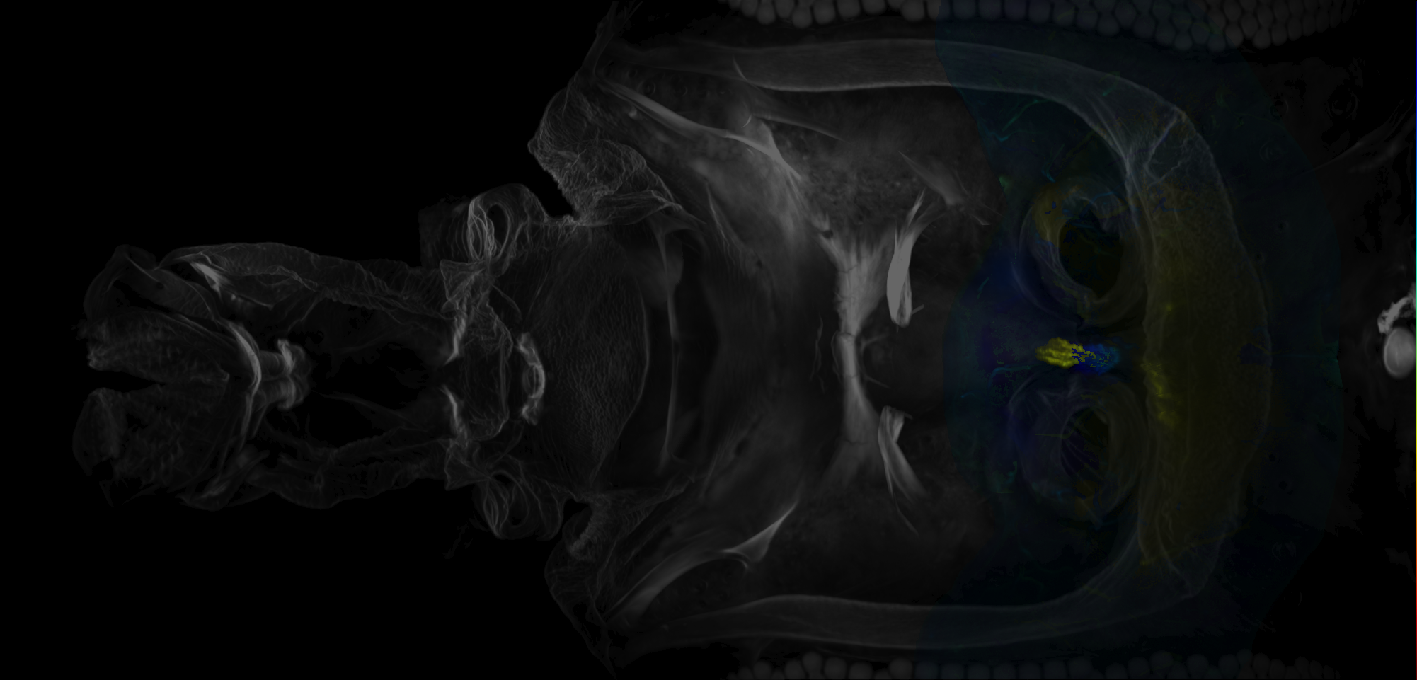


Images of proboscis muscles from McKellar et al., 2020
ID: HeadMusclesMcKellar2020
Images of proboscis muscles from McKellar et al., 2020
Open in VFB 3D Browser →Imaging Technique
- confocal microscopy
Downloads
Image files aligned to adult brain on Adult Head (McKellar2020):
Image files aligned to adult pharynx on Adult Head (McKellar2020):
Image files aligned to proboscis muscle 12 ventral on Adult Head (McKellar2020):
Image files aligned to proboscis muscle 4 on Adult Head (McKellar2020):
Image files aligned to proboscis muscle 3 medial and lateral fibers on Adult Head (McKellar2020):
Image files aligned to proboscis muscle 1 on Adult Head (McKellar2020):
Image files aligned to proboscis muscle 8 on Adult Head (McKellar2020):
Image files aligned to proboscis muscle 12 dorsal on Adult Head (McKellar2020):
Image files aligned to proboscis muscle 11 ventral on Adult Head (McKellar2020):
Image files aligned to proboscis muscle 10 on Adult Head (McKellar2020):
References
66 - Ito lab adult brain lineage clone image set [Ito2013]


Ito lab adult brain lineage clone image set
ID: Ito2013
An exhaustive set of lineage clones covering the adult brain from Kei Ito's lab.
Open in VFB 3D Browser →Imaging Technique
- confocal microscopy
Downloads
Image files aligned to VPNp&v1 clone of Ito 2013:
Image files aligned to VLPl&p1 clone of Ito 2013:
Image files aligned to SLPpm1 clone of Ito 2013:
Image files aligned to LHa3 clone of Ito 2013:
Image files aligned to SLPpm4 clone of Ito 2013:
Image files aligned to VPNp&v1 clone of Ito 2013:
Image files aligned to VLPl&p1 clone of Ito 2013:
Image files aligned to SLPpm1 clone of Ito 2013:
Image files aligned to LHa3 clone of Ito 2013:
Image files aligned to SLPpm4 clone of Ito 2013:
References
67 - JRC 2018 templates & ROIs [JRC2018]


JRC 2018 templates & ROIs
ID: JRC2018
JRC2018 templates with regions of interest painted by Kazunori Shinomiya
Open in VFB 3D Browser →Imaging Technique
- computer graphic
Downloads
Image files aligned to GA on JRC2018Unisex adult brain:
Image files aligned to FLA on JRC2018Unisex adult brain:
Image files aligned to CRE on JRC2018Unisex adult brain:
Image files aligned to WED on JRC2018Unisex adult brain:
Image files aligned to PLP on JRC2018Unisex adult brain:
Image files aligned to AOTU on JRC2018Unisex adult brain:
Image files aligned to FB on JRC2018Unisex adult brain:
Image files aligned to bL on JRC2018Unisex adult brain:
Image files aligned to CA on JRC2018Unisex adult brain:
Image files aligned to AME on JRC2018Unisex adult brain:
Images in dataset JRC 2018 templates & ROIs (46 total)
| Thumbnail | Name | Tags |
|---|---|---|
| [GA on JRC2018Unisex adult brain](/reports/VFB_00102281) | Nervous_system, Adult, Synaptic_neuropil_domain |
| [FLA on JRC2018Unisex adult brain](/reports/VFB_00102274) | Nervous_system, Adult, Synaptic_neuropil_domain |
| [CRE on JRC2018Unisex adult brain](/reports/VFB_00102171) | Nervous_system, Adult, Synaptic_neuropil_domain |
| [WED on JRC2018Unisex adult brain](/reports/VFB_00102154) | Nervous_system, Adult, Synaptic_neuropil_domain |
 | [PLP on JRC2018Unisex adult brain](/reports/VFB_00102152) | Nervous_system, Adult, Synaptic_neuropil_domain |
 | [AOTU on JRC2018Unisex adult brain](/reports/VFB_00102141) | Nervous_system, Adult, Synaptic_neuropil_domain |
 | [FB on JRC2018Unisex adult brain](/reports/VFB_00102134) | Nervous_system, Adult, Synaptic_neuropil_domain |
 | [bL on JRC2018Unisex adult brain](/reports/VFB_00102123) | Nervous_system, Adult, Synaptic_neuropil_subdomain |
 | [CA on JRC2018Unisex adult brain](/reports/VFB_00102114) | Nervous_system, Adult, Synaptic_neuropil_domain |
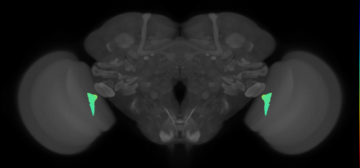 | [AME on JRC2018Unisex adult brain](/reports/VFB_00102108) | Nervous_system, Adult, Visual_system, Synaptic_neuropil_subdomain |
References
68 - JRC_FlyEM_Hemibrain neurons Version 1.0.1 [Xu2020Neurons]
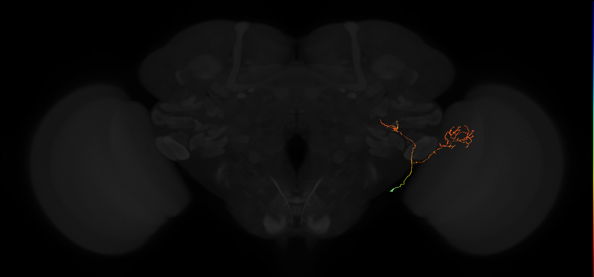
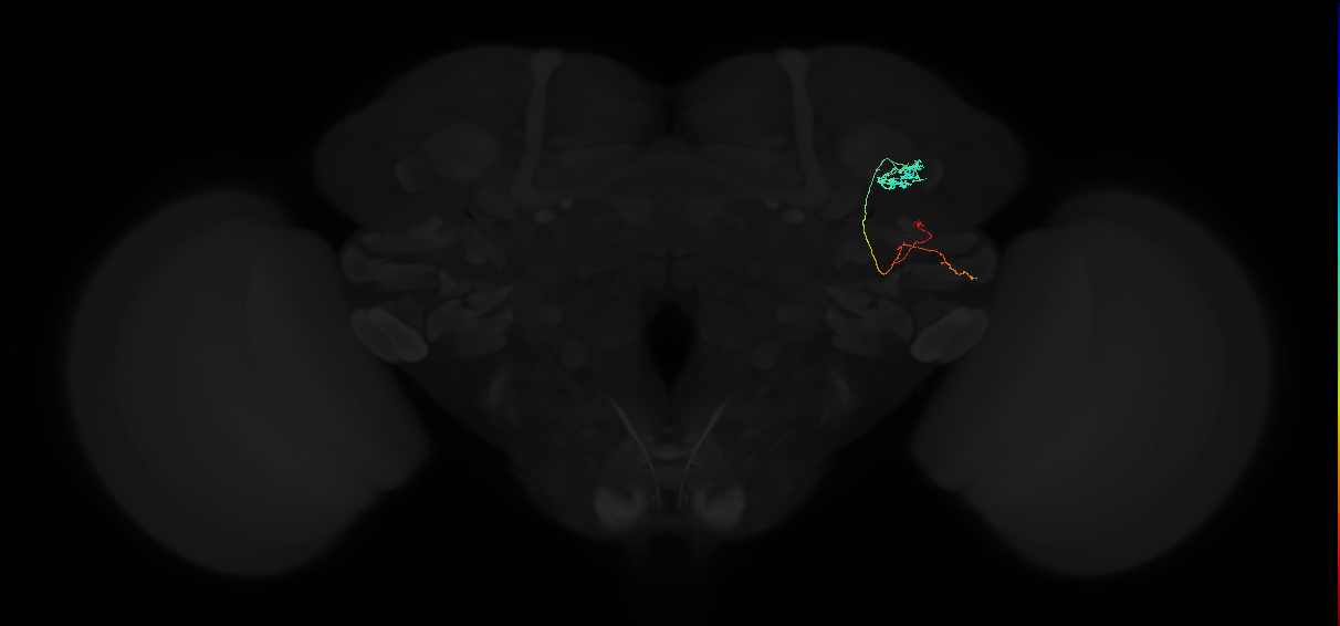
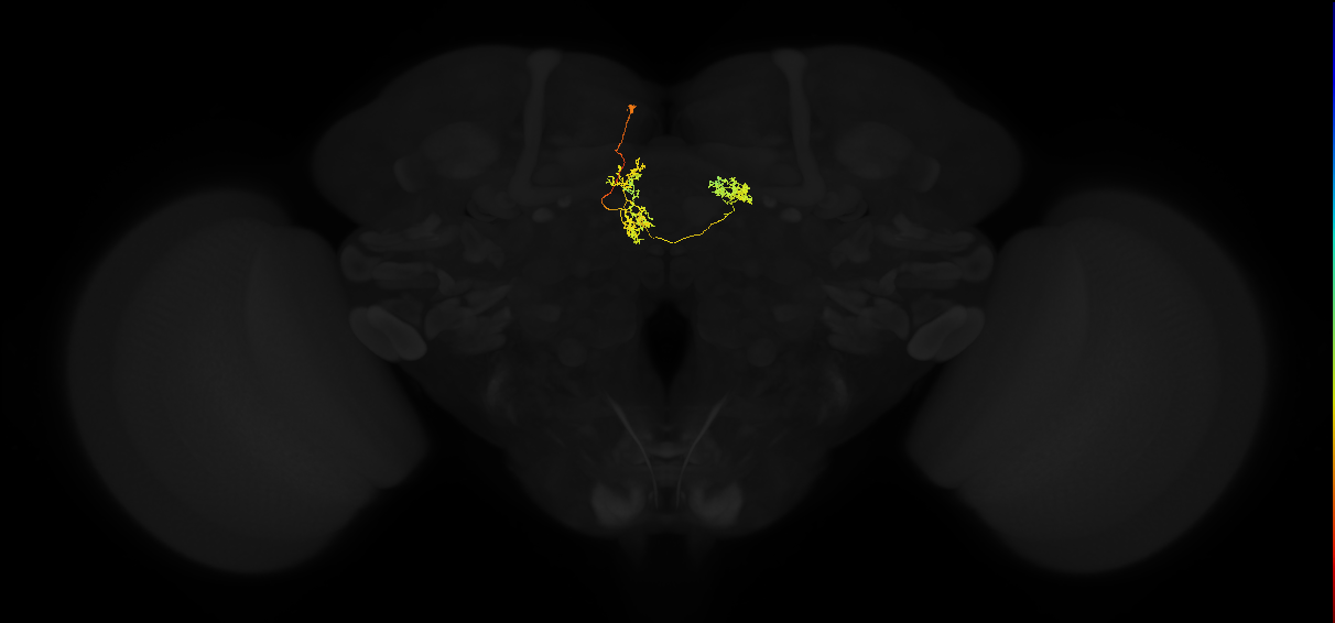
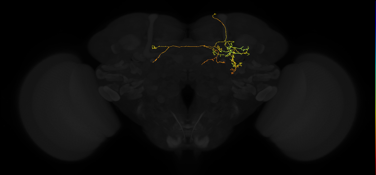
JRC_FlyEM_Hemibrain neurons Version 1.0.1
ID: Xu2020Neurons
Version 1.0.1 of reconstruction JRC_FlyEM_Hemibrain neurons and their synaptic connectivity
Open in VFB 3D Browser →Cross References
Imaging Technique
- focussed ion beam scanning electron microscopy (FIB-SEM)
Downloads
Image files aligned to 1529933124:
Image files aligned to 1077122311:
Image files aligned to 1165542823:
Image files aligned to 418970102:
Image files aligned to 363716362:
Image files aligned to 1529933124:
Image files aligned to 1077122311:
Image files aligned to 1165542823:
Image files aligned to 418970102:
Image files aligned to 363716362:
References
69 - JRC_FlyEM_Hemibrain neurons Version 1.2.1 [Xu2020NeuronsV1point2point1]
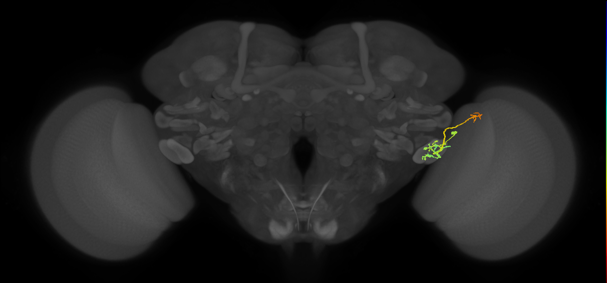


JRC_FlyEM_Hemibrain neurons Version 1.2.1
ID: Xu2020NeuronsV1point2point1
Version 1.2.1 of reconstruction JRC_FlyEM_Hemibrain neurons and their synaptic connectivity
Open in VFB 3D Browser →Cross References
Imaging Technique
- focussed ion beam scanning electron microscopy (FIB-SEM)
- serial block face SEM (SBFSEM)
Downloads
Image files aligned to LC17:
Image files aligned to KCg-m_R:
Image files aligned to AVLP379_R:
Image files aligned to AVLP176_d_R:
Image files aligned to DM6_adPN_L:
Image files aligned to LC17:
Image files aligned to KCg-m_R:
Image files aligned to IB003_R:
Image files aligned to AVLP379_R:
Image files aligned to AVLP176_d_R:
References
70 - JRC_FlyEM_Hemibrain painted domains [Xu2020roi]
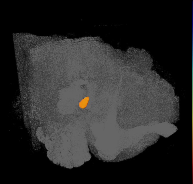


JRC_FlyEM_Hemibrain painted domains
ID: Xu2020roi
Synapse segmentation and regions of interest on JRC_FlyEM_Hemibrain
Open in VFB 3D Browser →Cross References
- JRC_FlyEM_Hemibrain painted domains on JRC FlyEM Project (hemibrain)
- JRC_FlyEM_Hemibrain painted domains on Neuprint web interface - hemibrain:v1.0.1
Imaging Technique
- computer graphic
Downloads
Image files aligned to NO2(L) on JRC_FlyEM_Hemibrain:
Image files aligned to NO1(R) on JRC_FlyEM_Hemibrain:
Image files aligned to VES(L) on JRC_FlyEM_Hemibrain:
Image files aligned to PB(L6) on JRC_FlyEM_Hemibrain:
Image files aligned to GA(R) on JRC_FlyEM_Hemibrain:
Image files aligned to AL(L) on JRC_FlyEM_Hemibrain:
Image files aligned to AOT(R) on JRC_FlyEM_Hemibrain:
Image files aligned to EB on JRC_FlyEM_Hemibrain:
Image files aligned to FBl9 on JRC_FlyEM_Hemibrain:
Image files aligned to EBr5 on JRC_FlyEM_Hemibrain:
References
71 - L3 Larval CNS Template (Truman2016) [Truman2016]
L3 Larval CNS Template (Truman2016)
ID: Truman2016
Template image by James W. Truman and the Fly Light Project Team at HHMI Janelia Research Campus.
Open in VFB 3D Browser →72 - L3 neuropils (WoodHartenstein2018) [WoodHartenstein2018]


Imaging Technique
- computer graphic
Downloads
Image files aligned to left ventromedial neuropil of A4 on L3 CNS template, Wood2018:
Image files aligned to left centromedial neuropil of A3 on L3 CNS template, Wood2018:
Image files aligned to left centromedial neuropil of maxillary segment on L3 CNS template, Wood2018:
Image files aligned to right ventromedial neuropil of A5 on L3 CNS template, Wood2018:
Image files aligned to right centromedial neuropil of A5 on L3 CNS template, Wood2018:
Image files aligned to left central neuropil of A5 on L3 CNS template, Wood2018:
Image files aligned to left dorsomedial neuropil of A6 on L3 CNS template, Wood2018:
Image files aligned to left ventrolateral neuropil of A3 on L3 CNS template, Wood2018:
Image files aligned to left ventrolateral subdivision of ventral nerve cord on L3 CNS template, Wood2018:
Image files aligned to left dorsolateral neuropil of A4 on L3 CNS template, Wood2018:
73 - larval hugin neurons - EM (Schlegel2016) [Schlegel2016]


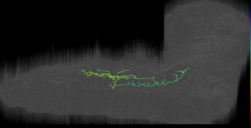
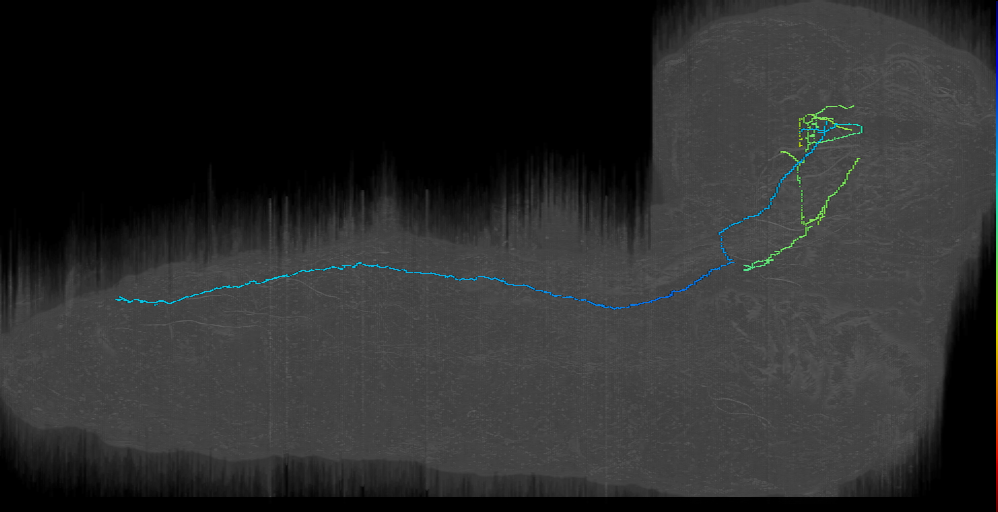
Cross References
Imaging Technique
- transmission electron microscopy (TEM)
Downloads
Image files aligned to AN-R-Sens-B1-AVa-20 (L1EM:16442344):
Image files aligned to AN-L-Sens-B1-ACa-02 (L1EM:13744425):
Image files aligned to post B5 17 l (L1EM:12824059):
Image files aligned to DPMm1 Brain descending into VNC left (L1EM:10716080):
Image files aligned to IPC 1 left (L1EM:10329208):
Image files aligned to v’td_a4l (Abdominal Sensory) (L1EM:9835169):
Image files aligned to v’td_a6l (Abdominal Sensory) (L1EM:7340629):
Image files aligned to AN-L-Sens-B1-ACa-01 (L1EM:2284021):
Image files aligned to Premotor for PaN + MN - paired with #10885848? (L1EM:2620299):
Image files aligned to AN-L-Sens-B1-AVa-21 (L1EM:1435466):
References
74 - Larval MB neurons - EM (Eichler2017) [Eichler2017]


Larval MB neurons - EM (Eichler2017)
ID: Eichler2017
Eichler, Li, Litwin-Kumar et al. 2017
Open in VFB 3D Browser →Cross References
Imaging Technique
- transmission electron microscopy (TEM)
Downloads
Image files aligned to KC (L1EM:5836163):
Image files aligned to KC (L1EM:9574259):
Image files aligned to MBON-d3 (L1EM:9074101):
Image files aligned to KC (L1EM:8957325):
Image files aligned to MBON-q1 (L1EM:7910624):
Image files aligned to KC (L1EM:8177991):
Image files aligned to KC (L1EM:3819157):
Image files aligned to MBIN-b1 (L1EM:8689674):
Image files aligned to 3rd-order Thermo right; CPLd interneuron (L1EM:8598886):
Image files aligned to 63a PN (L1EM:8245513):
References
75 - Larval motor circuit neurons (Zwart2016) [Zwart2016]


Cross References
Imaging Technique
- transmission electron microscopy (TEM)
Downloads
Image files aligned to MN8 (RF:SBM) LT3_a1r (L1EM:4881629):
Image files aligned to A23a_a1r (L1EM:4729046):
Image files aligned to A03a4_a1r (L1EM:4543281):
Image files aligned to MN22/23 (RF:LT2/LT3) LT1_a1r (L1EM:4102728):
Image files aligned to A03a3_t3l (was T03a1) (L1EM:14242929):
Image files aligned to A19l_a1l (L1EM:5358138):
Image files aligned to A27n_a1r (L1EM:4124563):
Image files aligned to MN21/22 (RF:LT1/LT2) LT2_a1l (L1EM:6467481):
Image files aligned to MN22/23 (RF:LT2/LT3) LT1_a1l (L1EM:4042315):
Image files aligned to A03g_a1l (L1EM:5309932):
References
76 - Larval olfactory system neurons - EM (Berk2016) [Berck2016]
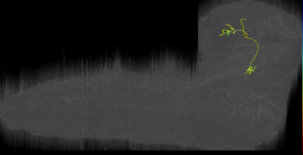


Larval olfactory system neurons - EM (Berk2016)
ID: Berck2016
Berck, Khandelwal et al. 2016
Open in VFB 3D Browser →Cross References
Imaging Technique
- transmission electron microscopy (TEM)
Downloads
Image files aligned to Giant-step left (L1EM:17844900):
Image files aligned to OAN-a1 (L1EM:17068730):
Image files aligned to mPN D (L1EM:6451332):
Image files aligned to picky 3 left (L1EM:8274021):
Image files aligned to 83a PN (L1EM:8243455):
Image files aligned to 35a ORN left (L1EM:3465659):
Image files aligned to mPN seahorse (L1EM:4324898):
Image files aligned to 45a ORN right (L1EM:3963891):
Image files aligned to 47a & 33b ORN right (L1EM:3896629):
Image files aligned to 45b ORN right (L1EM:3813723):
References
77 - Larval peristaltic locomotor system neurons - EM (Fushiki2016) [Fushiki2016]

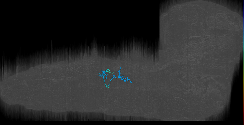


Larval peristaltic locomotor system neurons - EM (Fushiki2016)
ID: Fushiki2016
Cross References
Imaging Technique
- transmission electron microscopy (TEM)
Downloads
Image files aligned to A03EGB_a2r (EGHBAL) (L1EM:4848300):
Image files aligned to A27k_a2r (L1EM:4113612):
Image files aligned to A08s1_a1r (L1EM:14913093):
Image files aligned to A27a_a1r (L1EM:14046245):
Image files aligned to a03d/e_a2l (L1EM:12901480):
Image files aligned to A01a_a1l (L1EM:12140925):
Image files aligned to A14 dude?_a1r (L1EM:10133525):
Image files aligned to vdaC_a1r (class II) (L1EM:10053734):
Image files aligned to A08s1_a1l (L1EM:8059283):
Image files aligned to A05k_a1l (L1EM:4990880):
References
78 - larval sensorimotor decision pathways (Jovanic2016) [Jovanic2016]


larval sensorimotor decision pathways (Jovanic2016)
ID: Jovanic2016
Jovanic, Schneider-Mizell et al. 2016
Open in VFB 3D Browser →Cross References
Imaging Technique
- transmission electron microscopy (TEM)
Downloads
Image files aligned to Griddle-1 t3l (L1EM:9867541):
Image files aligned to Ladder-a_t3l (L1EM:8614658):
Image files aligned to Ladder-f a1 (L1EM:8577316):
Image files aligned to Drunken-1 a1l (L1EM:6207232):
Image files aligned to Griddle-1 a1r (L1EM:4824878):
Image files aligned to Ladder-d_a1 (L1EM:471705):
Image files aligned to Ladder-c a1 (L1EM:471720):
Image files aligned to chos_lch5-1_a1 (L1EM:7764785):
Image files aligned to chos_v’ch_a1 (L1EM:7628068):
Image files aligned to Basin3_a1 (L1EM:3091943):
References
79 - larval visual circuit neurons (Larderet2017) [Larderet2017]


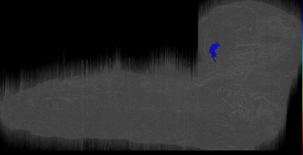
larval visual circuit neurons (Larderet2017)
ID: Larderet2017
Larderet, Fritsch et al. 2017
Open in VFB 3D Browser →Cross References
Imaging Technique
- transmission electron microscopy (TEM)
Downloads
Image files aligned to anlagen cell_left (L1EM:8449457):
Image files aligned to anlagen cell_left (L1EM:8448809):
Image files aligned to nc2;right (L1EM:8270683):
Image files aligned to OLP4;right (L1EM:8480418):
Image files aligned to Rh6PR-6;left (L1EM:2216131):
Image files aligned to Rh6PR-8;right (L1EM:946481):
Image files aligned to Rh6PR-3;right (L1EM:902038):
Image files aligned to Rh5PR-1;right (L1EM:766246):
Image files aligned to Rh5PR-5;right (L1EM:7568455):
Image files aligned to Vision PN 1 (L1EM:8111135):
References
80 - Larval wave neurons and circuit partners - EM (Takagi2017) [Takagi2017]


Larval wave neurons and circuit partners - EM (Takagi2017)
ID: Takagi2017
Cross References
Imaging Technique
- transmission electron microscopy (TEM)
Downloads
Image files aligned to MN30 RP4_a1l (L1EM:14194234):
Image files aligned to Pre-goro6 left (L1EM:18694051):
Image files aligned to A02o_a1r Wave-1 (L1EM:18684928):
Image files aligned to ddac t2r (L1EM:18303100):
Image files aligned to swallowtail t2l (L1EM:18189977):
Image files aligned to T19v_t1l (L1EM:16605707):
Image files aligned to MN2? ISN MN (U3?-MN2- NB7-1?)_a2r (L1EM:13875211):
Image files aligned to goro-like t1r (L1EM:6724973):
Image files aligned to MN1 aCC_a3l (L1EM:3931434):
Image files aligned to Pre-goro6 right (L1EM:360861):
References
81 - Lee lab adult brain lineage clone image set [Yu2013]


Lee lab adult brain lineage clone image set
ID: Yu2013
An exhaustive set of lineage clones covering the adult brain from Tzumin Lee's lab.
Open in VFB 3D Browser →Imaging Technique
- confocal microscopy
Downloads
Image files aligned to LHl3 clone of Yu 2013:
Image files aligned to CREa1 (male) clone of Yu 2013:
Image files aligned to SLPal1 clone of Yu 2013:
Image files aligned to VLPa1 clone of Yu 2013:
Image files aligned to CREa1 (female) clone of Yu 201:
Image files aligned to LHl3 clone of Yu 2013:
Image files aligned to CREa1 (male) clone of Yu 2013:
Image files aligned to SLPal1 clone of Yu 2013:
Image files aligned to VLPa1 clone of Yu 2013:
Image files aligned to CREa1 (female) clone of Yu 201:
References
82 - LexA driver targetting mechanosensory eye bristles (Hampel2017) [Hampel2017]
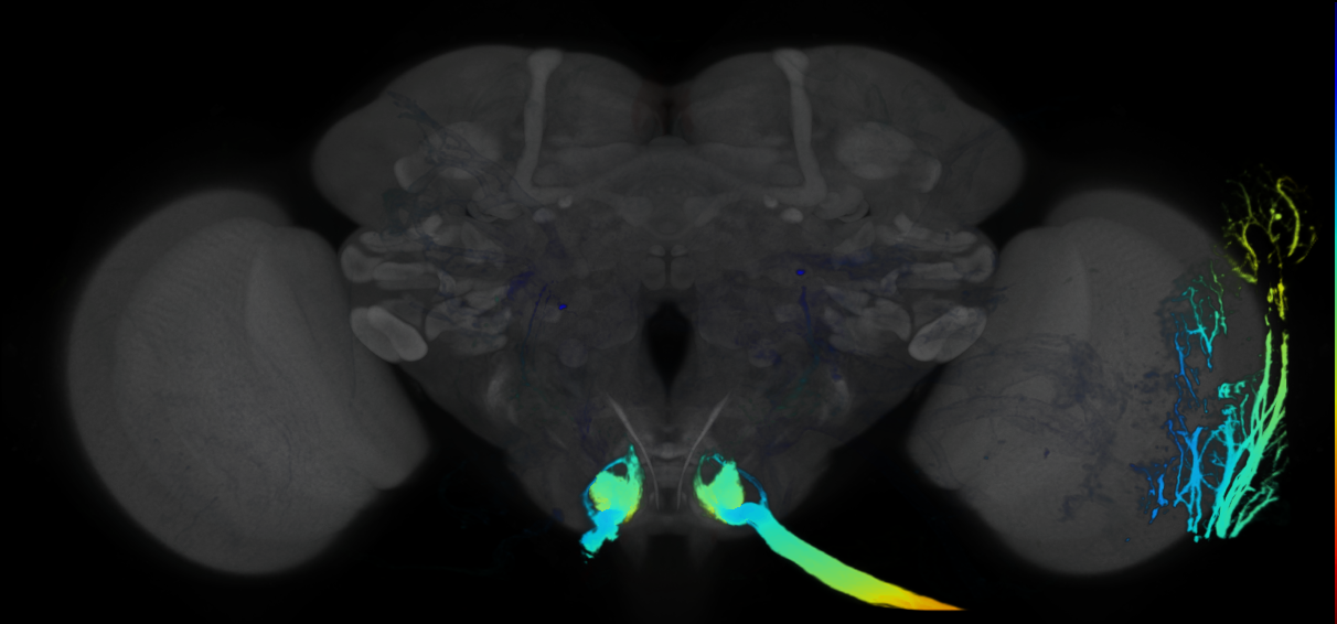

LexA driver targetting mechanosensory eye bristles (Hampel2017)
ID: Hampel2017
Simultaneous activation of parallel sensory pathways promotes a grooming sequence in Drosophila
Open in VFB 3D Browser →Imaging Technique
- confocal microscopy
Downloads
Image files aligned to VT17251 LexA that targets mechanosensory eye bristles:
Image files aligned to VT17251 LexA that targets mechanosensory eye bristles:
References
83 - Male Adult Nerve Cord (MANC) connectome neurons [Takemura2023]


Male Adult Nerve Cord (MANC) connectome neurons
ID: Takemura2023
Version 1.2.1 of the Male Adult Nerve Cord (MANC) connectome. Includes neuron skeletons, meshes and their synaptic connectivity
Open in VFB 3D Browser →Cross References
- Male Adult Nerve Cord (MANC) connectome neurons on JRC FlyEM Project (manc-connectome)
Imaging Technique
- focussed ion beam scanning electron microscopy (FIB-SEM)
Downloads
Image files aligned to SNxx06_AbNT_R (MANC:171382):
Image files aligned to SNxx24_MesoLN_R (MANC:153827):
Image files aligned to SAxxxx_MesoLN_L (MANC:151730):
Image files aligned to SNta04_ADMN_L (MANC:40198):
Image files aligned to TBD (MANC:32312):
Image files aligned to IN06A062_T3_R (MANC:28627):
Image files aligned to IN01B010_T2_L (MANC:28341):
Image files aligned to SAppxx_DMetaN_R (MANC:26975):
Image files aligned to AN07B077_A1_R (MANC:24154):
Image files aligned to IN17A045_T2_R (MANC:16561):
Images in dataset Male Adult Nerve Cord (MANC) connectome neurons (23759 total)
| Thumbnail | Name | Tags |
|---|---|---|
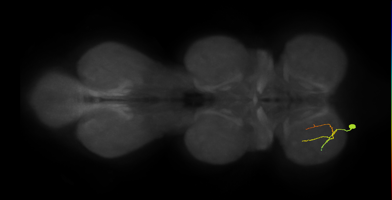 | [TBD (MANC:166374)](/reports/VFB_jrcv3kdi) | secondary_neuron, Motor_neuron, Glutamatergic, Adult, lineage_15_NB2-3 |
| [TBD (MANC:166374)](/reports/VFB_jrcv3kdi) | secondary_neuron, Motor_neuron, Glutamatergic, Adult, lineage_15_NB2-3 |
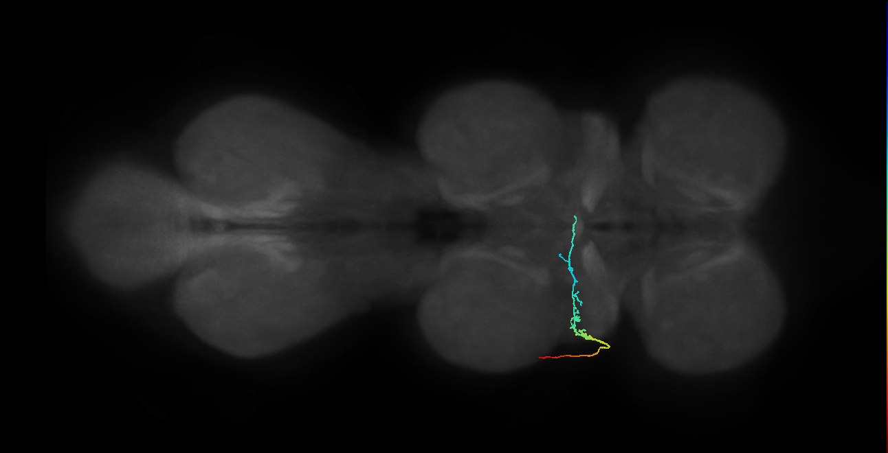 | [SNta09_PDMN_L (MANC:29754)](/reports/VFB_jrcv0myi) | Nervous_system, Mechanosensory_system, Sensory_neuron, Adult, Cholinergic |
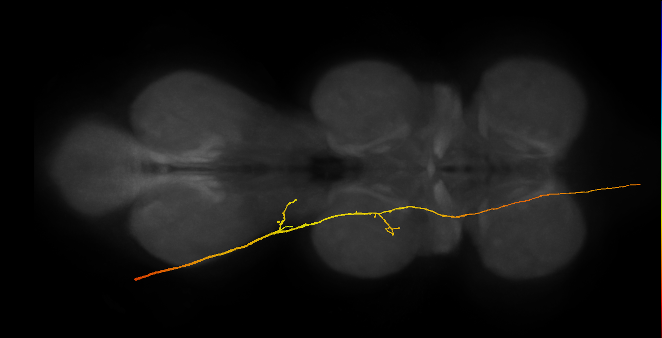 | [SAppxx_DMetaN_L (MANC:18718)](/reports/VFB_jrcv0efy) | Nervous_system, Sensory_neuron, Mechanosensory_system, Adult, Cholinergic |
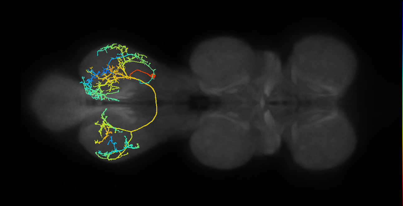 | [IN19B025_T3_R (MANC:11843)](/reports/VFB_jrcv094z) | secondary_neuron, Adult, Cholinergic, lineage_19_NB6-2 |
 | [IN09A002_T3_R (MANC:10380)](/reports/VFB_jrcv080c) | primary_neuron, lineage_9_NB3-5, GABAergic, Adult |
| [IN09A002_T3_R (MANC:10380)](/reports/VFB_jrcv080c) | primary_neuron, lineage_9_NB3-5, GABAergic, Adult |
View all 23759 results in VFB →
References
84 - male-cns [Berg2025]
male-cns
ID: Berg2025
Neurons from male-cns - Berg et al. (2025). Includes neuron skeletons, meshes and their synaptic connectivity.
Open in VFB 3D Browser →Imaging Technique
- serial block face SEM (SBFSEM)
Downloads
Image files aligned to PVLP072_R (MaleCNS:17920):
Image files aligned to KCg-m_R (MaleCNS:65161):
Image files aligned to LPT101_R (MaleCNS:76560):
Image files aligned to Mi15_L (MaleCNS:160866):
Image files aligned to PVLP072_R (MaleCNS:17920):
Image files aligned to PS359_R (MaleCNS:11266):
Image files aligned to R8y_L (MaleCNS:241537):
Image files aligned to Tm3_L (MaleCNS:90864):
Image files aligned to Tm20_L (MaleCNS:72642):
Image files aligned to Tm33_L (MaleCNS:147429):
List all available images of male-cns (10 total)
| Thumbnail | Name | Tags |
|---|---|---|
| LPT101_R (MaleCNS:76560) | Nervous_system, Adult, Visual_system |
| PVLP072_R (MaleCNS:17920) | Nervous_system, Adult |
 | PVLP072_R (MaleCNS:17920) | Nervous_system, Adult |
 | Tm20_L (MaleCNS:72642) | Nervous_system, Adult, Visual_system |
 | Tm3_L (MaleCNS:90864) | Nervous_system, Adult, Visual_system |
Images in dataset male-cns (210775 total)
| Thumbnail | Name | Tags |
|---|---|---|
 | [KCg-m_R (MaleCNS:65161)](/reports/VFB_jrmc2kkc) | Peptidergic, lineage_MBp2, Adult, Cholinergic, Dopaminergic |
 | [LPT101_R (MaleCNS:76560)](/reports/VFB_jrmc23z3) | lineage_VLPp&l1_DPLpv, Visual_system, Adult, Cholinergic |
 | [PVLP072_R (MaleCNS:17920)](/reports/VFB_jrmc1kb7) | Adult, lineage_VLPl&p1_BLVp2, Cholinergic |
 | [PVLP072_R (MaleCNS:17920)](/reports/VFB_jrmc1kb7) | Adult, lineage_VLPl&p1_BLVp2, Cholinergic |
 | [Tm3_L (MaleCNS:90864)](/reports/VFB_jrmc0m7w) | secondary_neuron, Nervous_system, Visual_system, GABAergic, Adult, Cholinergic |
 | [Tm20_L (MaleCNS:72642)](/reports/VFB_jrmc0l0u) | secondary_neuron, Nervous_system, Adult, Visual_system, Cholinergic |
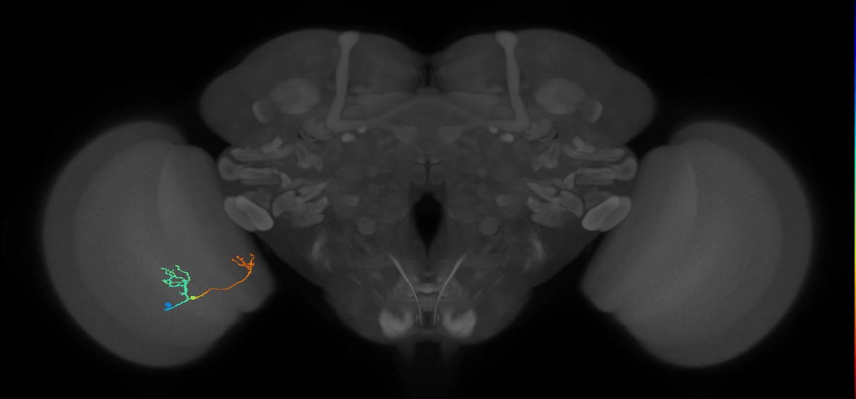 | [Tm33_L (MaleCNS:147429)](/reports/VFB_jrmc0gbp) | Nervous_system, Visual_system, Adult, Cholinergic |
View all 210775 results in VFB →
References
85 - MBONs and split-GAL4 lines that target them (Aso2014) [Aso2014]


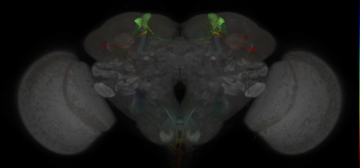
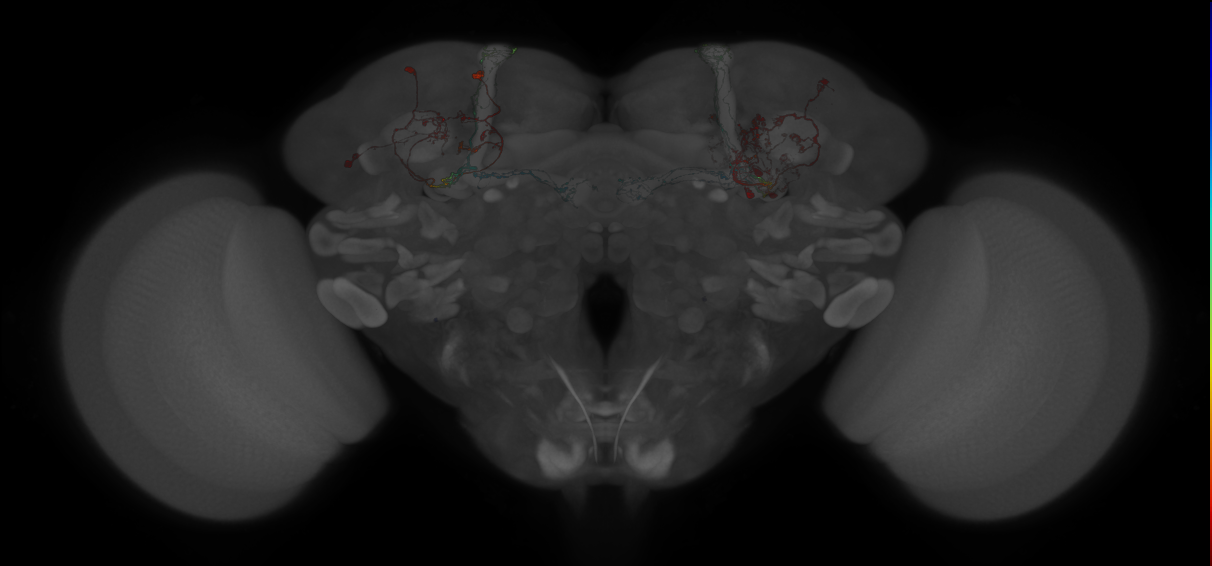
Imaging Technique
- confocal microscopy
Downloads
Image files aligned to MB194B:
Image files aligned to MB310C:
Image files aligned to JRC_MB304B_Brain_20170829_34_A5_63x:
Image files aligned to JRC_MB185B_Brain_20121205_19_F5_63x:
Image files aligned to JRC_MB301B_Brain_20121214_20_A2_20x:
Image files aligned to MB304B:
Image files aligned to MB018B:
Image files aligned to MB315C:
Image files aligned to MB109B:
Image files aligned to MB109B:
References
86 - MCFO images of GMR-GAL4 lines from Dionne et al., 2018 [Gen1MCFODionne2018]
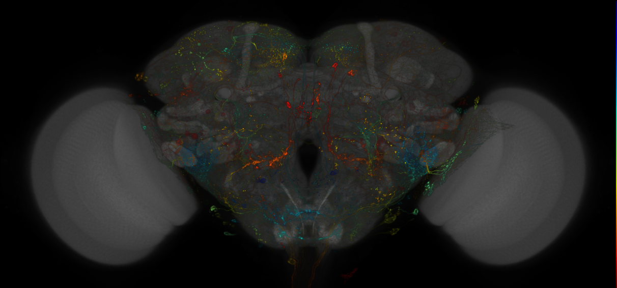
MCFO images of GMR-GAL4 lines from Dionne et al., 2018
ID: Gen1MCFODionne2018
MCFO images of GMR-GAL4 lines from Dionne et al., 2018
Open in VFB 3D Browser →Imaging Technique
- confocal microscopy
Downloads
Image files aligned to R9B08:
Image files aligned to R9B08:
Image files aligned to R71C03:
Image files aligned to R61E08:
Image files aligned to R59F08:
Image files aligned to R53H02:
Image files aligned to R11A03:
Image files aligned to R9B08:
Image files aligned to R75A04:
Image files aligned to R59F08:
References
87 - MCFO images of GMR-GAL4 lines from Jenett et al., 2012 [Gen1MCFOJenett2012]
MCFO images of GMR-GAL4 lines from Jenett et al., 2012
ID: Gen1MCFOJenett2012
MCFO images of GMR-GAL4 lines from Jenett et al., 2012
Open in VFB 3D Browser →Imaging Technique
- confocal microscopy
Downloads
Image files aligned to R74B06:
Image files aligned to R49A11:
Image files aligned to R51F11:
Image files aligned to R30A09:
Image files aligned to R17C11:
Image files aligned to R12C03:
Image files aligned to R70H05:
Image files aligned to R53D01:
Image files aligned to R32H08:
Image files aligned to R24B03:
References
88 - MCFO images of GMR-GAL4 lines from Jovanic et al., 2019 [Gen1MCFOJovanic2019]
MCFO images of GMR-GAL4 lines from Jovanic et al., 2019
ID: Gen1MCFOJovanic2019
MCFO images of GMR-GAL4 lines from Jovanic et al., 2019
Open in VFB 3D Browser →References
89 - MCFO images of GMR-GAL4 lines from Pfeiffer et al., 2010 [Gen1MCFOPfeiffer2010]


MCFO images of GMR-GAL4 lines from Pfeiffer et al., 2010
ID: Gen1MCFOPfeiffer2010
MCFO images of GMR-GAL4 lines from Pfeiffer et al., 2010
Open in VFB 3D Browser →Imaging Technique
- confocal microscopy
Downloads
Image files aligned to R22C03:
Image files aligned to R22C03:
Image files aligned to R22C03:
Image files aligned to R22C03:
References
90 - MCFO images of VT-GAL4 lines from Tirian et al., 2017 [Gen1MCFOTirian2017]
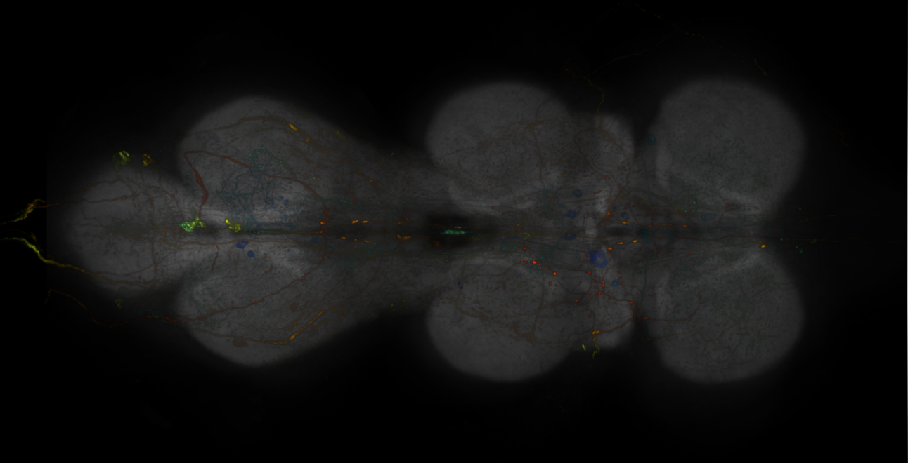
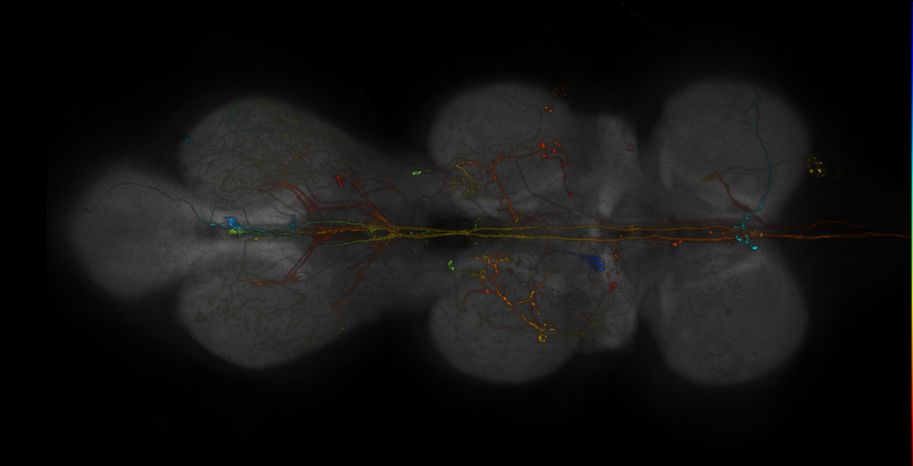
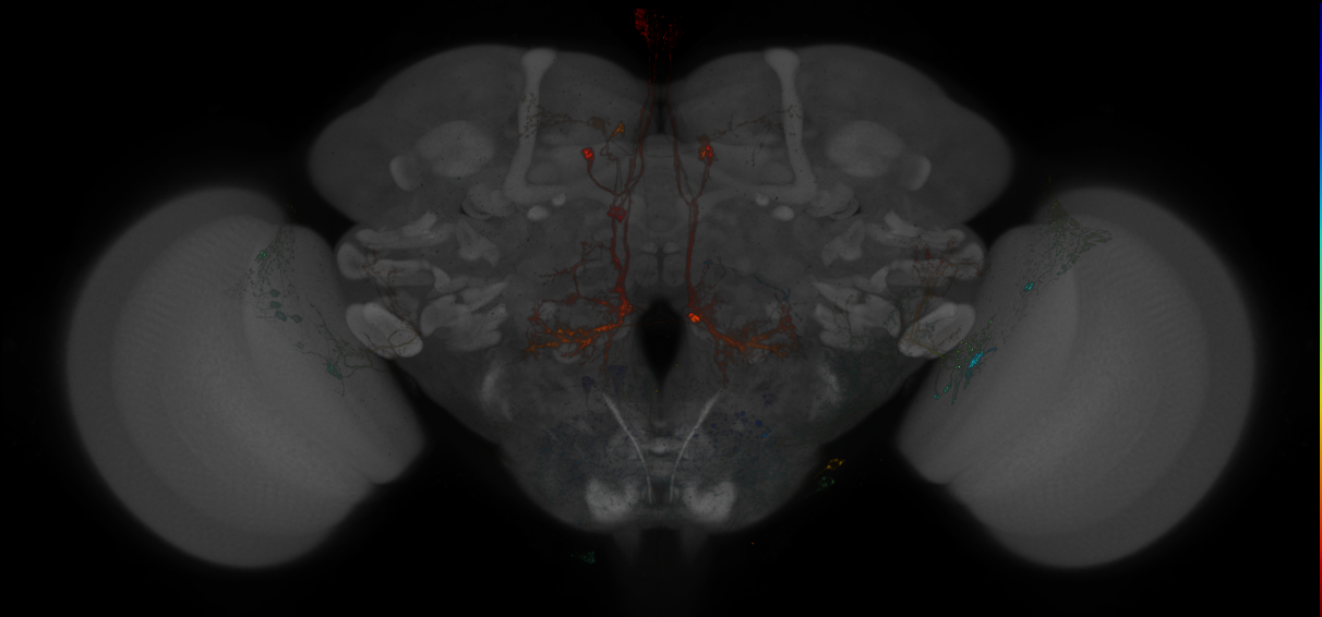
MCFO images of VT-GAL4 lines from Tirian et al., 2017
ID: Gen1MCFOTirian2017
MCFO images of VT-GAL4 lines from Tirian et al., 2017
Open in VFB 3D Browser →Imaging Technique
- confocal microscopy
Downloads
Image files aligned to VT013947:
Image files aligned to VT042478:
Image files aligned to VT005110:
Image files aligned to VT037554:
Image files aligned to VT026324:
Image files aligned to VT026846:
Image files aligned to VT060077:
Image files aligned to VT064919:
Image files aligned to VT021374:
Image files aligned to VT039961:
References
91 - Millimeter-scale imaging of a Drosophila leg at single-neuron resolution [Kuan2020]


Millimeter-scale imaging of a Drosophila leg at single-neuron resolution
ID: Kuan2020
Millimeter-scale imaging of a Drosophila leg at single-neuron resolution from Dense neuronal reconstruction through X-ray holographic nano-tomography (Kuan et al., 2020
Open in VFB 3D Browser →Cross References
Imaging Technique
- X-ray computed tomography
Downloads
Image files aligned to muscle 93997 (T1LEG:610138):
Image files aligned to muscle 93960 (T1LEG:610005):
Image files aligned to neuron 112385 (T1LEG:609978):
Image files aligned to neuron 146462 (T1LEG:607420):
Image files aligned to scolopidium 103301 (T1LEG:606244):
Image files aligned to neuron 74067 (T1LEG:605410):
Image files aligned to neuron 82535 (T1LEG:604381):
Image files aligned to muscle 94768 (T1LEG:604163):
Image files aligned to neuron 74033 (T1LEG:603032):
Image files aligned to neuron 88315 (T1LEG:602851):
References
92 - Neurons involved in courtship and song (Lillvis2018) [Lillvis2018]


Neurons involved in courtship and song (Lillvis2018)
ID: Lillvis2018
single example of pIP10 neuron in adult CNS
Open in VFB 3D Browser →Imaging Technique
- confocal microscopy
Downloads
Image files aligned to pIP10 neuron in the adult brain:
Image files aligned to pIP10 neuron in the adult brain:
Image files aligned to pIP10 neuron in the adult VNS:
References
93 - Neurons involved in larval fast escape response - EM (Ohyama2016) [Ohyama2015]


Neurons involved in larval fast escape response - EM (Ohyama2016)
ID: Ohyama2015
Ohyama, Schneider-Mizell et al. 2015
Open in VFB 3D Browser →Cross References
Imaging Technique
- transmission electron microscopy (TEM)
Downloads
Image files aligned to chos_vchAB2_a1 (L1EM:9771412):
Image files aligned to chos_vchAB1_a1 (L1EM:7746883):
Image files aligned to A23g_a1r (L1EM:5997121):
Image files aligned to T05t_t3 (L1EM:5343532):
Image files aligned to A12m_a1l (L1EM:1767828):
Image files aligned to T05t_t3 (L1EM:3957604):
Image files aligned to chos_lch5-24-1_a1 (L1EM:7577773):
Image files aligned to A00c_a6 (L1EM:1690576):
Image files aligned to Basin-1 a4r (L1EM:1398260):
Image files aligned to chos_lch5-24-1_a1 (L1EM:162293):
References
94 - Nociceptive circuit neurons - EM (Gerhard2017) [Gerhard2017]


Cross References
Imaging Technique
- transmission electron microscopy (TEM)
Downloads
Image files aligned to fragment (L1EM:18322843):
Image files aligned to A09l_t3r Down-and-back-1 (L1EM:10053642):
Image files aligned to A00c_A8l? (L1EM:5731368):
Image files aligned to A09q_a7r classIV-related projection D right (L1EM:734837):
Image files aligned to A09l_t3l Down-and-back-1 (L1EM:7124056):
Image files aligned to A09o_a3r (L1EM:2693651):
Image files aligned to A09e_a3 (L1EM:14795310):
Image files aligned to ppk_v’ada_a1 (L1EM:3786734):
Image files aligned to Basin2_a1 (L1EM:3074106):
Image files aligned to Basin4_a1 (L1EM:3040481):
References
95 - Nociceptive system neurons - EM (Burgos2017) [Burgos2018]


Nociceptive system neurons - EM (Burgos2017)
ID: Burgos2018
Burgos et al 2018
Open in VFB 3D Browser →Cross References
Imaging Technique
- transmission electron microscopy (TEM)
Downloads
Image files aligned to A03g_a1r (L1EM:10855206):
Image files aligned to A02o_a5r Wave-1 (L1EM:1401061):
Image files aligned to A02o_a5l Wave-1 (L1EM:3437872):
Image files aligned to A02o_a6l Wave-1 (L1EM:754774):
Image files aligned to ddaA a1l class III md (L1EM:722935):
Image files aligned to A09l_a1l (Down-and-back-1) (L1EM:12468812):
Image files aligned to A09e_a4 (L1EM:627858):
Image files aligned to A02o_a3r Wave-1 (L1EM:12946335):
Image files aligned to gorogoro t2l (L1EM:3720037):
Image files aligned to A09c_a4l Basin-4 (L1EM:587083):
References
96 - Optic Lobe connectome neurons from optic_lobe:v1.0.1, Nern et al., 2024 [Nern2024]
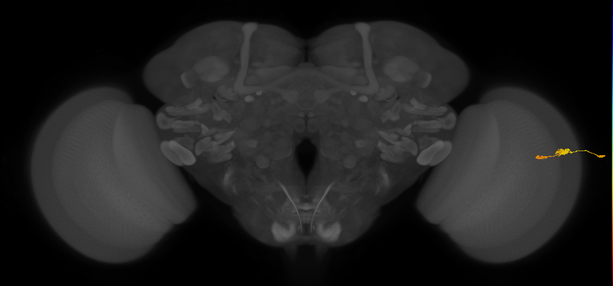
Optic Lobe connectome neurons from optic_lobe:v1.0.1, Nern et al., 2024
ID: Nern2024
Optic Lobe connectome neurons from optic_lobe:v1.0.1, Nern et al., 2024. Includes neuron skeletons, meshes and their synaptic connectivity. These optic lobe neurons are traced from part of the Male CNS EM volume
Open in VFB 3D Browser →Cross References
- Optic Lobe connectome neurons from optic_lobe:v1.0.1, Nern et al., 2024 on JRC FlyEM Project (optic-lobe)
Imaging Technique
- focussed ion beam scanning electron microscopy (FIB-SEM)
Downloads
Image files aligned to TmY14_R (JRC_OpticLobe:21055):
Image files aligned to Tm9_R (JRC_OpticLobe:139720):
Image files aligned to Pm7_R (JRC_OpticLobe:13462):
Image files aligned to Mi9_R (JRC_OpticLobe:75757):
Image files aligned to Mi2_R (JRC_OpticLobe:123558):
Image files aligned to Mi14_R (JRC_OpticLobe:66688):
Image files aligned to Li25_R (JRC_OpticLobe:29128):
Image files aligned to LC21_R (JRC_OpticLobe:53170):
Image files aligned to L4_R (JRC_OpticLobe:132535):
Image files aligned to L1_R (JRC_OpticLobe:33449):
References
97 - RNAseq_2020_Davis [PRJNA480794]
RNAseq_2020_Davis
ID: PRJNA480794
A study using TAPIN RNA sequencing to build high resolution transcriptomes from selected adult optic lobe neurons.
Open in VFB 3D Browser →Cross References
- RNAseq_2020_Davis on Gene Expression Omnibus (GSE116969)
- RNAseq_2020_Davis on EMBL-EBI Expression Atlas (E-GEOD-116969)
References
98 - scRNAseq_2018_Davie [FBlc0006090]
scRNAseq_2018_Davie
ID: FBlc0006090
Single-cell RNA-seq study of the aging brain
Open in VFB 3D Browser →Cross References
References
99 - scRNAseq_2019_Avalos [FBlc0005362]
scRNAseq_2019_Avalos
ID: FBlc0005362
Single-cell RNA-seq study of first instar larval brains upon starvation
Open in VFB 3D Browser →Cross References
- scRNAseq_2019_Avalos on EMBL-EBI Single Cell Expression Atlas (E-GEOD-134722)
References
100 - scRNAseq_2020_Allen [FBlc0005603]
scRNAseq_2020_Allen
ID: FBlc0005603
Single-cell RNA-seq study of adult ventral nerve cords
Open in VFB 3D Browser →Cross References
- scRNAseq_2020_Allen on EMBL-EBI Single Cell Expression Atlas (E-GEOD-141807)
References
101 - scRNAseq_2020_Hormann [FBlc0006305]
scRNAseq_2020_Hormann
ID: FBlc0006305
Single-cell RNA-seq study of visual motion-sensing neurons
Open in VFB 3D Browser →Cross References
- scRNAseq_2020_Hormann on EMBL-EBI Single Cell Expression Atlas (E-GEOD-147987)
References
102 - scRNAseq_2020_Kurmangaliyev [FBlc0006237]
scRNAseq_2020_Kurmangaliyev
ID: FBlc0006237
Single-cell RNA-seq study of the pupal optic lobe
Open in VFB 3D Browser →Cross References
References
103 - scRNAseq_2021_Baker_CTRL [FBlc0005515]
scRNAseq_2021_Baker_CTRL
ID: FBlc0005515
Single-cell RNA-seq study of adult brain without cocaine exposure
Open in VFB 3D Browser →Cross References
References
104 - scRNAseq_2021_IngSimmons [FBlc0006191]
scRNAseq_2021_IngSimmons
ID: FBlc0006191
Single-cell RNA-seq study of gastrulating embryos
Open in VFB 3D Browser →Cross References
References
105 - scRNAseq_2021_Mokashi_CTRL [FBlc0005420]
scRNAseq_2021_Mokashi_CTRL
ID: FBlc0005420
Single-cell RNA-seq study of adult brain without alcohol exposure
Open in VFB 3D Browser →Cross References
References
106 - scRNAseq_2021_Ozel [FBlc0005659]
scRNAseq_2021_Ozel
ID: FBlc0005659
Single-cell RNA-seq study of pupal and adult optic lobes
Open in VFB 3D Browser →Cross References
References
107 - scRNAseq_2022_Calderon [FBlc0007797]
scRNAseq_2022_Calderon
ID: FBlc0007797
The continuum of Drosophila embryonic development at single-cell resolution
Open in VFB 3D Browser →Cross References
References
108 - scRNAseq_2022_FCA_FEMALE [FBlc0003846]
scRNAseq_2022_FCA_FEMALE
ID: FBlc0003846
Single-nucleus RNA-seq on cells from 5-days old female flies
Open in VFB 3D Browser →Cross References
References
109 - scRNAseq_2022_FCA_MALE [FBlc0004307]
scRNAseq_2022_FCA_MALE
ID: FBlc0004307
Single-nucleus RNA-seq on cells from 5-days old male flies
Open in VFB 3D Browser →Cross References
References
110 - scRNAseq_2022_FCA_MIXED [FBlc0004785]
scRNAseq_2022_FCA_MIXED
ID: FBlc0004785
Single-nucleus RNA-seq on cells from 5-days old female and male flies
Open in VFB 3D Browser →Cross References
References
111 - scRNAseq_2022_Konstantinides [FBlc0006404]
scRNAseq_2022_Konstantinides
ID: FBlc0006404
Single-cell RNA-seq study of larval optic lobes
Open in VFB 3D Browser →Cross References
References
112 - scRNAseq_2023_AFCA_D30 [FBlc0006423]
scRNAseq_2023_AFCA_D30
ID: FBlc0006423
Single-nucleus RNA-seq on cells from 30-days old flies
Open in VFB 3D Browser →Cross References
- scRNAseq_2023_AFCA_D30 on EMBL-EBI Single Cell Expression Atlas (E-GEOD-218661)
References
113 - scRNAseq_2023_AFCA_D30_FEMALE [FBlc0006424]
scRNAseq_2023_AFCA_D30_FEMALE
ID: FBlc0006424
Single-nucleus RNA-seq on cells from 30-days old female flies
Open in VFB 3D Browser →Cross References
References
114 - scRNAseq_2023_AFCA_D30_MALE [FBlc0006611]
scRNAseq_2023_AFCA_D30_MALE
ID: FBlc0006611
Single-nucleus RNA-seq on cells from 30-days old male flies
Open in VFB 3D Browser →Cross References
References
115 - scRNAseq_2023_AFCA_D50 [FBlc0006887]
scRNAseq_2023_AFCA_D50
ID: FBlc0006887
Single-nucleus RNA-seq on cells from 50-days old flies
Open in VFB 3D Browser →Cross References
- scRNAseq_2023_AFCA_D50 on EMBL-EBI Single Cell Expression Atlas (E-GEOD-218661)
References
116 - scRNAseq_2023_AFCA_D50_FEMALE [FBlc0006888]
scRNAseq_2023_AFCA_D50_FEMALE
ID: FBlc0006888
Single-nucleus RNA-seq on cells from 50-days old female flies
Open in VFB 3D Browser →Cross References
References
117 - scRNAseq_2023_AFCA_D50_MALE [FBlc0007071]
scRNAseq_2023_AFCA_D50_MALE
ID: FBlc0007071
Single-nucleus RNA-seq on cells from 50-days old male flies
Open in VFB 3D Browser →Cross References
References
118 - scRNAseq_2023_AFCA_D70 [FBlc0007347]
scRNAseq_2023_AFCA_D70
ID: FBlc0007347
Single-nucleus RNA-seq on cells from 70-days old flies
Open in VFB 3D Browser →Cross References
- scRNAseq_2023_AFCA_D70 on EMBL-EBI Single Cell Expression Atlas (E-GEOD-218661)
References
119 - scRNAseq_2023_AFCA_D70_FEMALE [FBlc0007348]
scRNAseq_2023_AFCA_D70_FEMALE
ID: FBlc0007348
Single-nucleus RNA-seq on cells from 70-days old female flies
Open in VFB 3D Browser →Cross References
References
120 - scRNAseq_2023_AFCA_D70_MALE [FBlc0007532]
scRNAseq_2023_AFCA_D70_MALE
ID: FBlc0007532
Single-nucleus RNA-seq on cells from 70-days old male flies
Open in VFB 3D Browser →Cross References
References
121 - scRNAseq_2023_Saavedra [FBlc0006361]
scRNAseq_2023_Saavedra
ID: FBlc0006361
Single-cell RNA-seq study of the adult thorax
Open in VFB 3D Browser →Cross References
- scRNAseq_2023_Saavedra on EMBL-EBI Single Cell Expression Atlas (E-GEOD-227038)
References
122 - Split GAL4 lines for dopaminergic neurons, Xie2018 [Xie2018]


Imaging Technique
- confocal microscopy
Downloads
Image files aligned to P{R76F02-GAL4.DBD} ∩ P{ple-p65.AD.TH-D} expression pattern of Xie2018:
Image files aligned to P{R61H03-GAL4.DBD} ∩ P{R55C10-p65.AD} expression pattern of Xie2018:
Image files aligned to P{R76F02-GAL4.DBD} ∩ P{R60F07-p65.AD} expression pattern of Xie2018:
Image files aligned to P{R76F01-GAL4.DBD} ∩ P{ple-p65.AD.TH-D} expression pattern of Xie2018:
Image files aligned to P{R60F07-GAL4.DBD} ∩ P{R76F02-p65.AD} expression pattern of Xie2018:
Image files aligned to P{R76F02-GAL4.DBD} ∩ P{ple-p65.AD.TH-D} expression pattern of Xie2018:
Image files aligned to P{R61H03-GAL4.DBD} ∩ P{R55C10-p65.AD} expression pattern of Xie2018:
Image files aligned to P{R76F02-GAL4.DBD} ∩ P{R60F07-p65.AD} expression pattern of Xie2018:
Image files aligned to P{R76F01-GAL4.DBD} ∩ P{ple-p65.AD.TH-D} expression pattern of Xie2018:
Image files aligned to P{R60F07-GAL4.DBD} ∩ P{R76F02-p65.AD} expression pattern of Xie2018:
References
123 - split-GAL4 lines for EB neurons (Robie2017) [Robie2017]


split-GAL4 lines for EB neurons (Robie2017)
ID: Robie2017
split-GAL4 lines for Ellipsoid Body neurons from the (walking-more) brain-behavior map
Open in VFB 3D Browser →Imaging Technique
- confocal microscopy
Downloads
Image files aligned to SS02768:
Image files aligned to SS02764:
Image files aligned to SS02712:
Image files aligned to SS02709:
Image files aligned to SS02714:
Image files aligned to SS02710:
Image files aligned to SS02768:
Image files aligned to SS02768:
Image files aligned to SS02769:
Image files aligned to SS02767:
References
124 - split-GAL4 lines for descending neurons (Namiki2018) [FlyLight2019Namiki2018]


split-GAL4 lines for descending neurons (Namiki2018)
ID: FlyLight2019Namiki2018
Descending sensory-motor pathways in adult CNS
Open in VFB 3D Browser →Imaging Technique
- confocal microscopy
Downloads
Image files aligned to SS01572:
Image files aligned to SS01543:
Image files aligned to SS02633:
Image files aligned to SS02392:
Image files aligned to SS02542:
Image files aligned to SS02285:
Image files aligned to SS01590:
Image files aligned to SS01593:
Image files aligned to SS01552:
Image files aligned to SS00730:
References
125 - split-GAL4 lines for descending neurons (Namiki2018) [Namiki2018]


split-GAL4 lines for descending neurons (Namiki2018)
ID: Namiki2018
Descending sensory-motor pathways in adult CNS
Open in VFB 3D Browser →Imaging Technique
- confocal microscopy
Downloads
Image files aligned to SS01565:
Image files aligned to SS01056:
Image files aligned to SS02542:
Image files aligned to SS02276:
Image files aligned to SS01567:
Image files aligned to SS01549:
Image files aligned to SS02276:
Image files aligned to JRC_SS31976_VNC_20170307_32_G1_20x:
Image files aligned to SS02383:
Image files aligned to SS02276:
References
126 - split-GAL4 lines for dopaminergic neurons (AsoRubin2016) [AsoRubin2016]


split-GAL4 lines for dopaminergic neurons (AsoRubin2016)
ID: AsoRubin2016
For comparison of the properties of memories formed by optogenetic activation of individual DAN cell types
Open in VFB 3D Browser →Imaging Technique
- confocal microscopy
Downloads
Image files aligned to MB320C:
Image files aligned to MB320C:
Image files aligned to MB630B:
Image files aligned to MB320C:
Image files aligned to MB296B:
Image files aligned to MB213B:
Image files aligned to MB099C:
Image files aligned to MB630B:
Image files aligned to MB320C:
Image files aligned to MB099C:
References
127 - split-GAL4 lines for dopaminergic neurons (AsoRubin2016) [FlyLight2019AsoRubin2016]

split-GAL4 lines for dopaminergic neurons (AsoRubin2016)
ID: FlyLight2019AsoRubin2016
For comparison of the properties of memories formed by optogenetic activation of individual DAN cell types
Open in VFB 3D Browser →Imaging Technique
- confocal microscopy
Downloads
Image files aligned to MB630B:
References
128 - split-GAL4 lines for EB neurons (Robie2017) [FlyLight2019Robie2017]

split-GAL4 lines for EB neurons (Robie2017)
ID: FlyLight2019Robie2017
split-GAL4 lines for Ellipsoid Body neurons from the (walking-more) brain-behavior map
Open in VFB 3D Browser →Imaging Technique
- confocal microscopy
Downloads
Image files aligned to SS02763:
References
129 - split-GAL4 lines for LC VPNs (Wu2016) [FlyLight2019Wu2016]


split-GAL4 lines for LC VPNs (Wu2016)
ID: FlyLight2019Wu2016
split-GAL4 lines for the Lobula Columnar (LC) visual projection neurons (LC Paper)
Open in VFB 3D Browser →Imaging Technique
- confocal microscopy
Downloads
Image files aligned to SS02669:
Image files aligned to SS02645:
Image files aligned to OL0011B:
Image files aligned to OL0007B:
Image files aligned to OL0003B:
Image files aligned to OL0046B:
Image files aligned to OL0042B:
Image files aligned to OL0070B:
Image files aligned to SS00315:
Image files aligned to SS02669:
References
130 - split-GAL4 lines for LC VPNs (Wu2016) [Wu2016]
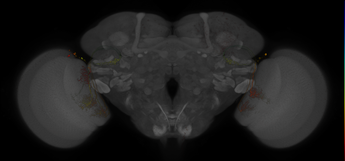


split-GAL4 lines for LC VPNs (Wu2016)
ID: Wu2016
split-GAL4 lines for the Lobula Columnar (LC) visual projection neurons (LC Paper)
Open in VFB 3D Browser →Imaging Technique
- confocal microscopy
Downloads
Image files aligned to OL0070B:
Image files aligned to OL0027B:
Image files aligned to OL0019B:
Image files aligned to SS00315:
Image files aligned to OL0022B:
Image files aligned to OL0007B:
Image files aligned to OL0015B:
Image files aligned to SS00937:
Image files aligned to OL0027B:
Image files aligned to OL0007B:
References
131 - Split-GAL4 lines from Aso et al., 2014b [SplitAso2014b]
Split-GAL4 lines from Aso et al., 2014b
ID: SplitAso2014b
Split-GAL4 lines labelling mushroom body output neurons from Aso et al., 2014b
Open in VFB 3D Browser →References
132 - Split-GAL4 lines from Baker et al., 2022 [SplitBaker2022]
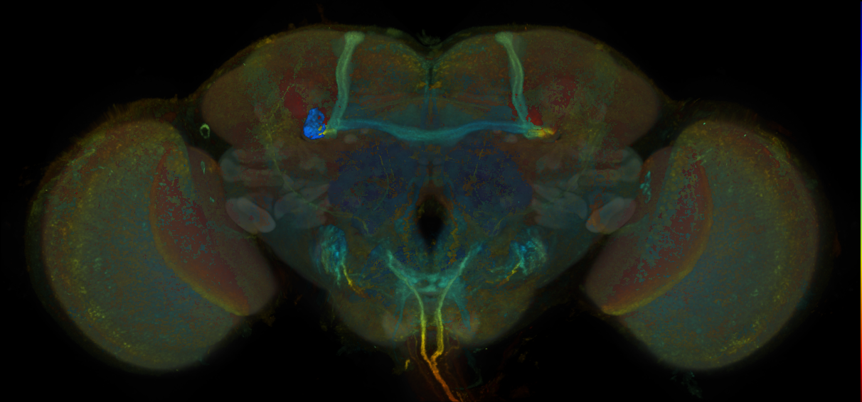
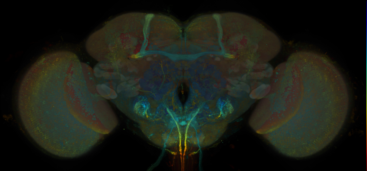
Split-GAL4 lines from Baker et al., 2022
ID: SplitBaker2022
Split-GAL4 lines from Baker et al., 2022
Open in VFB 3D Browser →Imaging Technique
- confocal microscopy
Downloads
Image files aligned to JRC_SS28822_Brain_20170712_31_A6_20x:
Image files aligned to JRC_SS28822_Brain_20170712_31_A4_20x:
Image files aligned to JRC_SS28822_Brain_20170712_31_A1_20x:
Image files aligned to JRC_SS36007_Brain_20170607_31_F5_20x:
Image files aligned to JRC_SS36007_Brain_20170607_31_F4_20x:
Image files aligned to JRC_SS36007_Brain_20170607_31_F3_20x:
Image files aligned to JRC_SS28822_Brain_20170712_31_A5_63x:
Image files aligned to JRC_SS36007_VNC_20170607_31_F2_20x:
References
133 - Split-GAL4 lines from Bidaye et al., 2014 [SplitBidaye2014]
Split-GAL4 lines from Bidaye et al., 2014
ID: SplitBidaye2014
Split-GAL4 lines from Bidaye et al., 2014
Open in VFB 3D Browser →References
134 - Split-GAL4 lines from Bidaye et al., 2020 [SplitBidaye2020]
Split-GAL4 lines from Bidaye et al., 2020
ID: SplitBidaye2020
Split-GAL4 lines from Bidaye et al., 2020
Open in VFB 3D Browser →135 - Split-GAL4 lines from Bogovic et al., 2020 [SplitBogovic2020]
Split-GAL4 lines from Bogovic et al., 2020
ID: SplitBogovic2020
Split-GAL4 lines used in the evaluation of JRC2018 templates from Bogovic et al., 2020
Open in VFB 3D Browser →Imaging Technique
- confocal microscopy
Downloads
Image files aligned to JRC_SS23208_VNC_20161102_32_E1_63x:
Image files aligned to JRC_SS23206_VNC_20161102_32_D2_63x:
Image files aligned to JRC_SS33017_VNC_20170223_32_E3_20x:
Image files aligned to JRC_SS33007_VNC_20170223_32_A3_20x:
Image files aligned to JRC_SS30777_VNC_20161220_32_C1_20x:
Image files aligned to JRC_SS30777_Brain_20161220_32_C5_20x:
Image files aligned to JRC_SS33007_Brain_20170714_46_G2_20x:
Image files aligned to JRC_SS32529_Brain_20170301_31_B3_20x:
Image files aligned to JRC_SS33017_Brain_20170223_32_E3_20x:
Image files aligned to JRC_SS33007_Brain_20170223_32_A6_20x:
References
136 - Split-GAL4 lines from Cheong et al., 2023 [SplitCheong2023]
Split-GAL4 lines from Cheong et al., 2023
ID: SplitCheong2023
Split-GAL4 lines from Cheong et al., 2023
Open in VFB 3D Browser →References
137 - Split-GAL4 lines from Cheong et al., 2024 [SplitCheong2024]
Split-GAL4 lines from Cheong et al., 2024
ID: SplitCheong2024
Split-GAL4 lines from Cheong et al., 2024
Open in VFB 3D Browser →References
138 - Split-GAL4 lines from Dag et al., 2019 [SplitDag2019]
Split-GAL4 lines from Dag et al., 2019
ID: SplitDag2019
Split-GAL4 lines from Dag et al., 2019
Open in VFB 3D Browser →References
139 - Split-GAL4 lines from Davis et al., 2020 [SplitDavis2020]


Split-GAL4 lines from Davis et al., 2020
ID: SplitDavis2020
Split-GAL4 lines from Davis et al 2020
Open in VFB 3D Browser →Imaging Technique
- confocal microscopy
Downloads
Image files aligned to SS02592:
Image files aligned to SS02700:
Image files aligned to SS02344:
Image files aligned to SS02427:
Image files aligned to SS02344:
Image files aligned to SS00657:
Image files aligned to SS00308:
Image files aligned to SS00309:
Image files aligned to SS03656:
Image files aligned to SS02448:
References
140 - Split-GAL4 lines from Ehrhardt et al., 2023 [SplitEhrhardt2023]
Split-GAL4 lines from Ehrhardt et al., 2023
ID: SplitEhrhardt2023
Split-GAL4 lines from Ehrhardt et al., 2023
Open in VFB 3D Browser →Imaging Technique
- confocal microscopy
Downloads
Image files aligned to JRC_SS41039_VNC_20170815_31_C1_20x:
Image files aligned to JRC_SS44028_Brain_20171020_23_A2_20x:
Image files aligned to JRC_SS44028_Brain_20171020_23_A1_20x:
References
141 - Split-GAL4 lines from Feng et al., 2014 [SplitFeng2014]
Split-GAL4 lines from Feng et al., 2014
ID: SplitFeng2014
Split-GAL4 lines from Feng et al., 2014
Open in VFB 3D Browser →References
142 - Split-GAL4 lines from Feng et al., 2020 [SplitFeng2020]
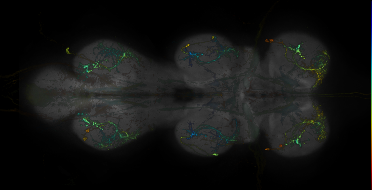
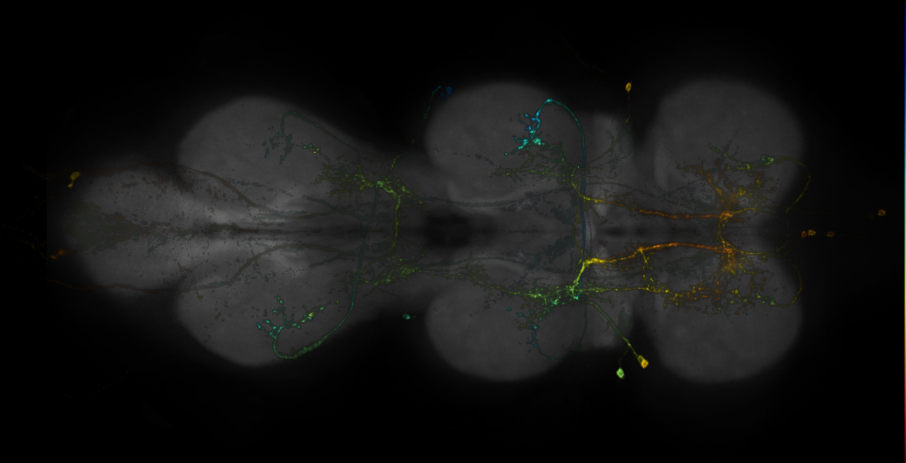
Split-GAL4 lines from Feng et al., 2020
ID: SplitFeng2020
Split-GAL4 lines labelling neurons downstream of Moonwalker Descending Neurons from Feng et al., 2020
Open in VFB 3D Browser →Imaging Technique
- confocal microscopy
Downloads
Image files aligned to JRC_SS27416_VNC_20171018_31_G4_63x:
Image files aligned to JRC_SS35560_VNC_20170912_32_F4_20x:
Image files aligned to JRC_SS32496_VNC_20170726_32_E1_20x:
Image files aligned to JRC_SS27416_VNC_20171018_31_G2_20x:
Image files aligned to JRC_SS33510_Brain_20170922_33_C4_20x:
Image files aligned to JRC_SS33542_Brain_20170912_32_G4_20x:
Image files aligned to JRC_SS33542_Brain_20170912_32_G3_20x:
Image files aligned to JRC_SS27416_Brain_20171018_31_G2_20x:
References
143 - Split-GAL4 lines from Gao et al., 2019 [SplitGao2019]
Split-GAL4 lines from Gao et al., 2019
ID: SplitGao2019
Split-GAL4 lines labelling PPM3 cluster subset neurons from Gao et al., 2019
Open in VFB 3D Browser →References
144 - Split-GAL4 lines from Garner et al., 2023 [SplitGarner2023]
Split-GAL4 lines from Garner et al., 2023
ID: SplitGarner2023
Split-GAL4 lines from Garner et al., 2023
Open in VFB 3D Browser →References
145 - Split-GAL4 lines from Giraldo et al., 2018 [SplitGiraldo2018]
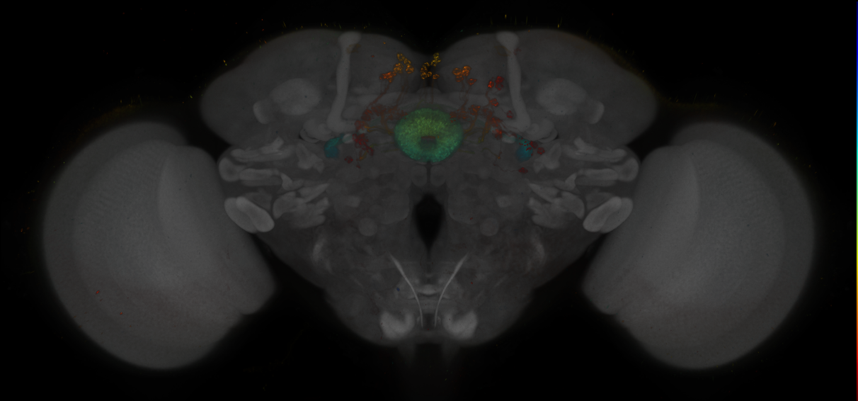
Split-GAL4 lines from Giraldo et al., 2018
ID: SplitGiraldo2018
Split-GAL4 lines from Giraldoet al 2018
Open in VFB 3D Browser →Imaging Technique
- confocal microscopy
Downloads
Image files aligned to SS00131:
Image files aligned to SS00131:
References
146 - Split-GAL4 lines from Gorko et al., 2024 [SplitGorko2024]
Split-GAL4 lines from Gorko et al., 2024
ID: SplitGorko2024
Split-GAL4 lines labelling neck motorneurons and proprioceptive neurons from Gorko et al., 2024
Open in VFB 3D Browser →References
147 - Split-GAL4 lines from Hattori et al., 2017 [SplitHattori2017]
Split-GAL4 lines from Hattori et al., 2017
ID: SplitHattori2017
Split-GAL4 lines labelling Mushroom Body neurons from Hattori et al 2017
Open in VFB 3D Browser →References
148 - Split-GAL4 lines from Hulse et al., 2021 [SplitHulse2021]
Split-GAL4 lines from Hulse et al., 2021
ID: SplitHulse2021
Split-GAL4 lines from Hulse et al., 2021
Open in VFB 3D Browser →References
149 - Split-GAL4 lines from Isaacson et al., 2023 [SplitIsaacson2023]
Split-GAL4 lines from Isaacson et al., 2023
ID: SplitIsaacson2023
Split-GAL4 lines from Isaacson et al., 2023
Open in VFB 3D Browser →References
150 - Split-GAL4 lines from Jovanic et al., 2019 [SplitJovanic2019]
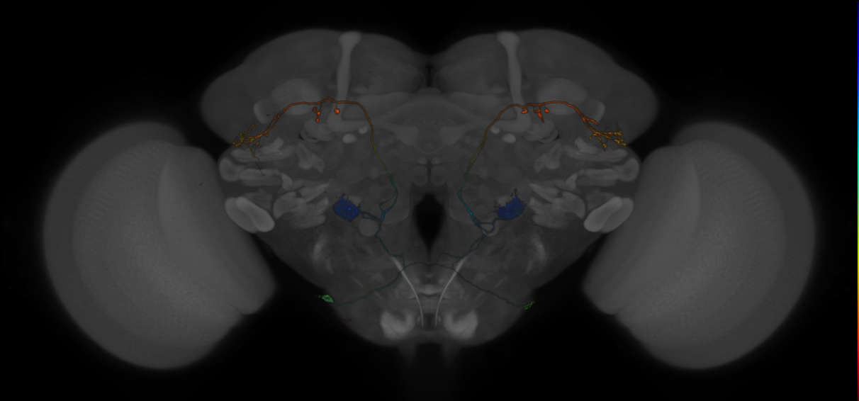
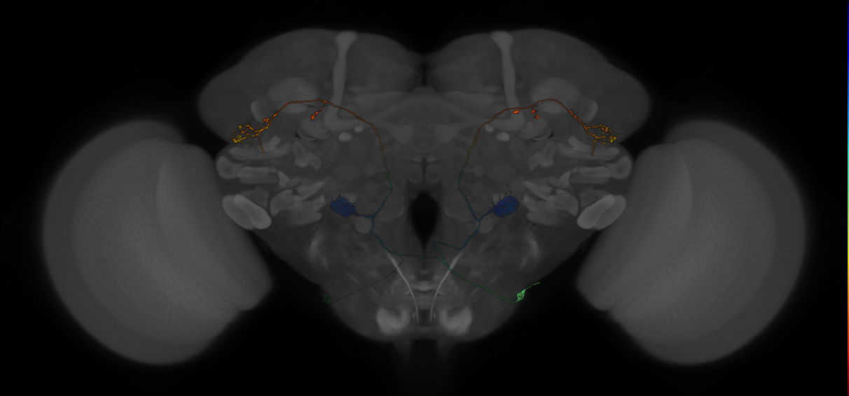
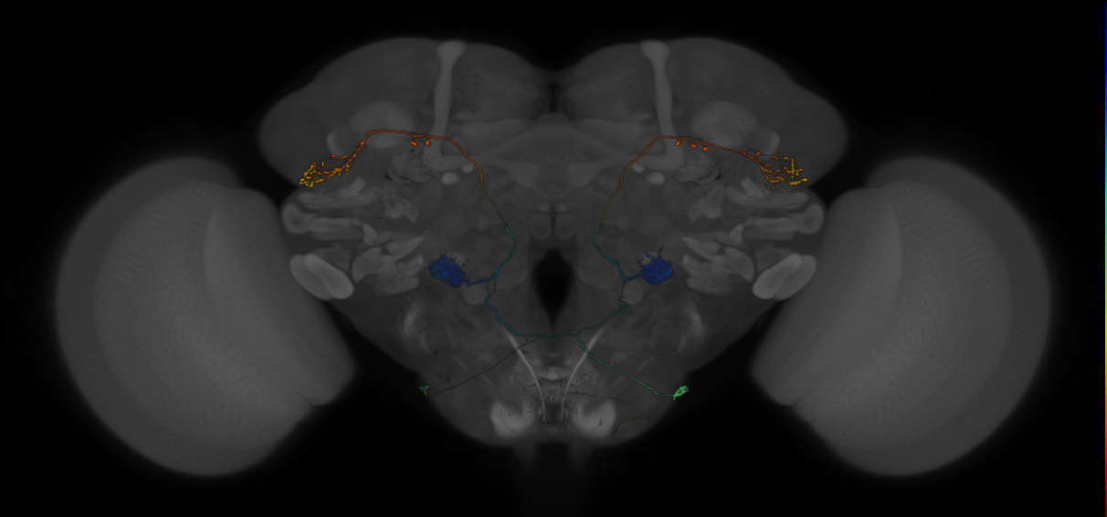
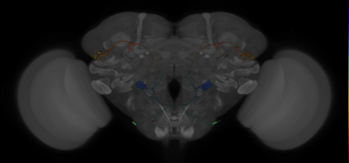
Split-GAL4 lines from Jovanic et al., 2019
ID: SplitJovanic2019
Split-GAL4 lines from Jovanic et al., 2019
Open in VFB 3D Browser →Imaging Technique
- confocal microscopy
Downloads
Image files aligned to JRC_MB546B_Brain_20121107_19_C2_63x:
Image files aligned to JRC_MB546B_Brain_20121107_19_D1_63x:
Image files aligned to JRC_MB546B_Brain_20121107_19_D3_20x:
Image files aligned to JRC_MB546B_Brain_20121107_19_D2_20x:
References
151 - Split-GAL4 lines from Kind et al., 2021 [SplitKind2021]
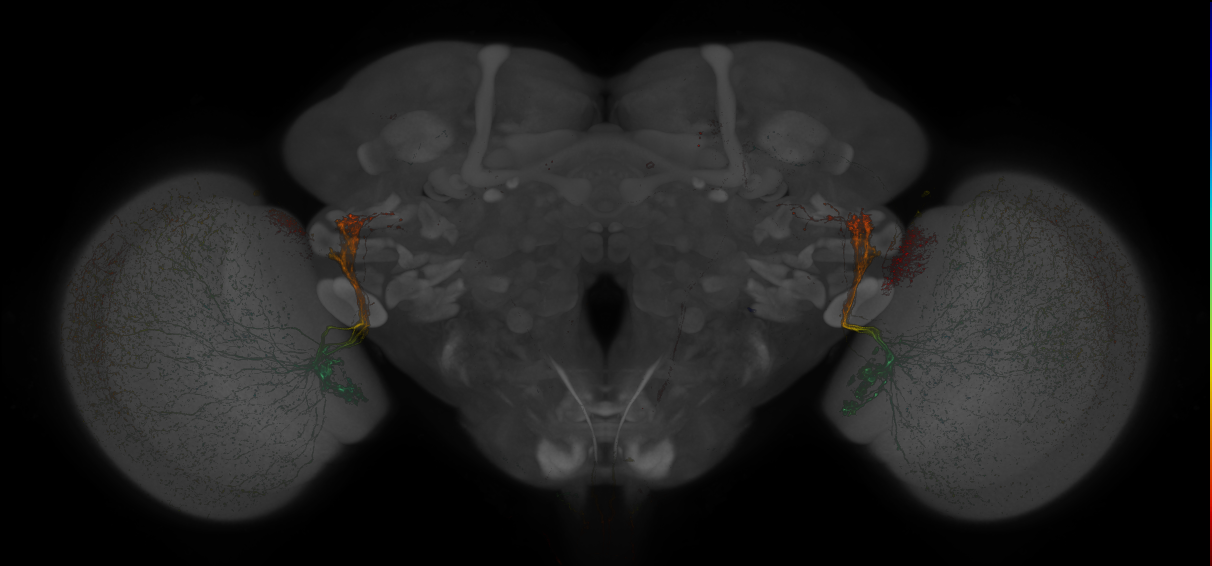
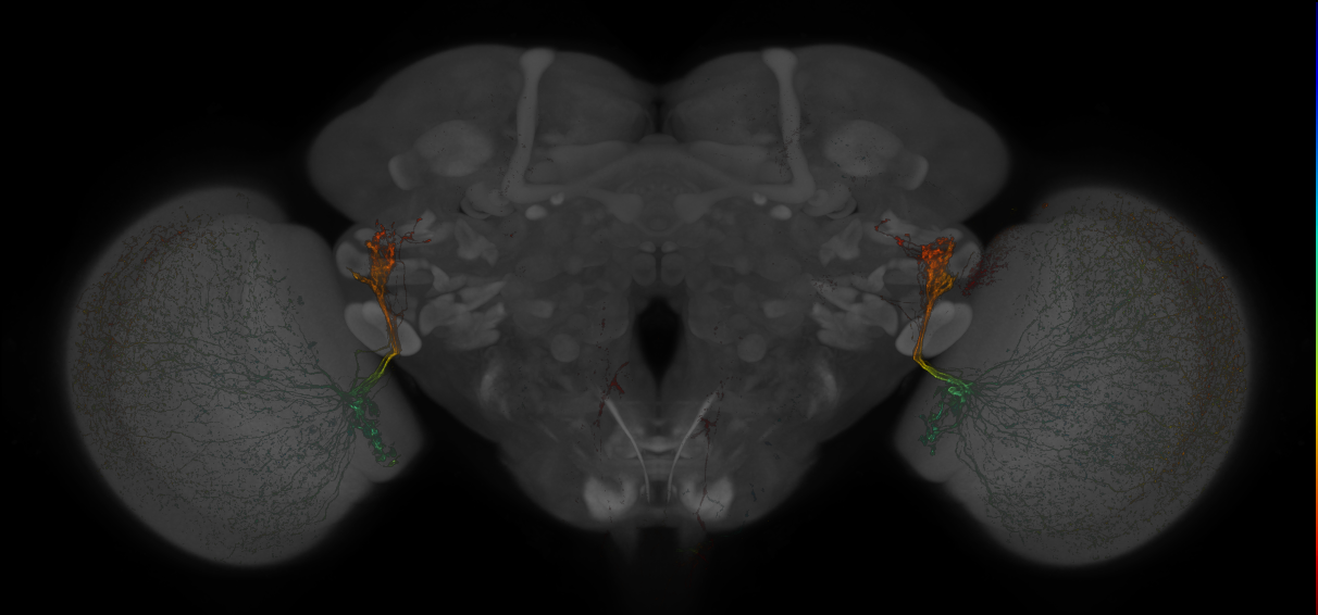
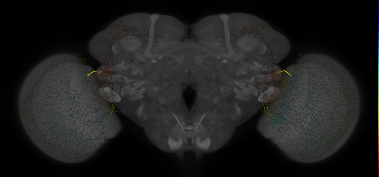
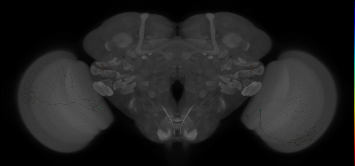
Split-GAL4 lines from Kind et al., 2021
ID: SplitKind2021
Split-GAL4 lines labelling R7 and R8 photoreceptor target neurons from Kind et al., 2021
Open in VFB 3D Browser →Imaging Technique
- confocal microscopy
Downloads
Image files aligned to JRC_SS28175_Brain_20161011_31_H2_63x:
Image files aligned to JRC_SS28175_Brain_20161011_31_H1_20x:
Image files aligned to JRC_SS02978_Brain_20150512_32_I3_20x:
Image files aligned to JRC_SS28175_Brain_20160909_28_E2_63x:
Image files aligned to JRC_SS02978_Brain_20150507_19_F8_20x:
Image files aligned to JRC_SS01015_Brain_20140115_19_B7_20x:
References
152 - Split-GAL4 lines from Klapoetke et al. 2017 [FlyLight2019Klapoetke2017]

Split-GAL4 lines from Klapoetke et al. 2017
ID: FlyLight2019Klapoetke2017
Split-GAL4 lines used for the characterization of LPLC2 anatomy and function
Open in VFB 3D Browser →Imaging Technique
- confocal microscopy
Downloads
Image files aligned to OL0047B:
References
153 - Split-GAL4 lines from Klapoetke et al. 2017 [Klapoetke2017]

Split-GAL4 lines from Klapoetke et al. 2017
ID: Klapoetke2017
Split-GAL4 lines used for the characterization of LPLC2 anatomy and function
Open in VFB 3D Browser →Imaging Technique
- confocal microscopy
Downloads
Image files aligned to SS00810:
Image files aligned to OL0047B:
Image files aligned to OL0047B:
Image files aligned to OL0047B:
Image files aligned to SS00810:
Image files aligned to OL0047B:
Image files aligned to SS00810:
Image files aligned to OL0047B:
Image files aligned to SS00810:
Image files aligned to OL0047B:
References
154 - Split-GAL4 lines from Klapoetke et al., 2022 [SplitKlapoetke2022]
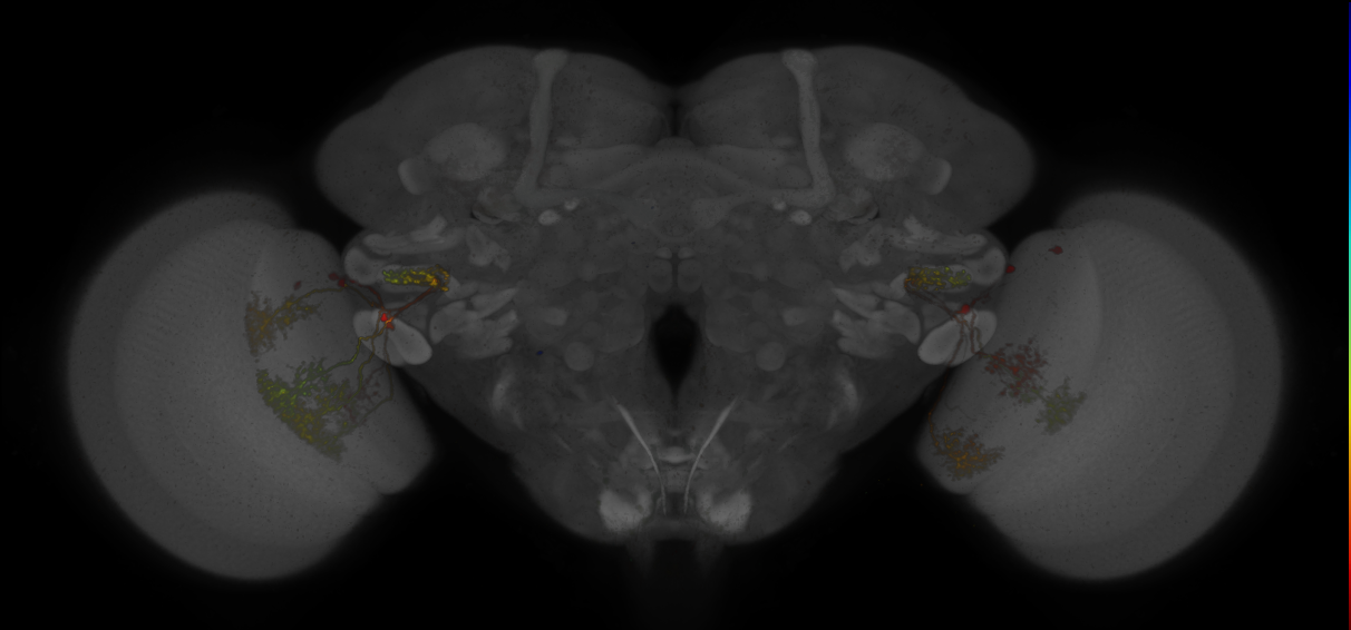
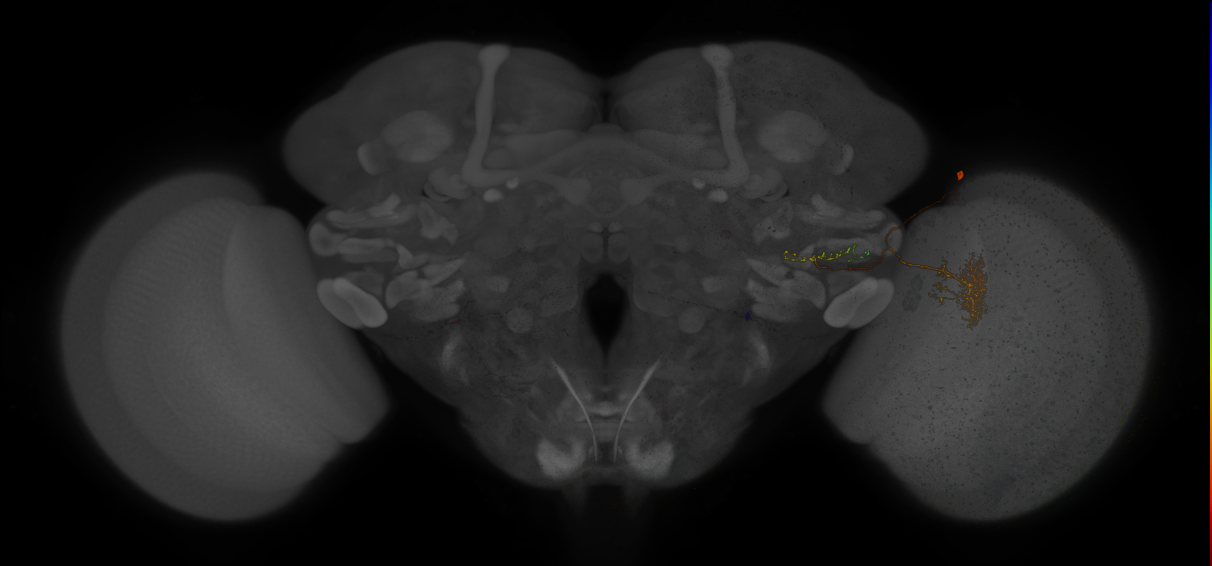
Split-GAL4 lines from Klapoetke et al., 2022
ID: SplitKlapoetke2022
Split-GAL4 lines labelling neurons for the characterization of a functionally ordered visual feature map from Klapoetke et al., 2022
Open in VFB 3D Browser →Imaging Technique
- confocal microscopy
Downloads
Image files aligned to JRC_SS17689_Brain_20160308_32_G2_63x:
Image files aligned to JRC_SS05037_Brain_20160311_22_B7_63x:
Image files aligned to JRC_OL0016B_Brain_20131125_33_F8_20x:
References
155 - Split-GAL4 lines from Liang et al., 2017 [SplitLiang2017]
Split-GAL4 lines from Liang et al., 2017
ID: SplitLiang2017
Split-GAL4 lines from Liang et al., 2017
Open in VFB 3D Browser →References
156 - Split-GAL4 lines from Lillvis et al., 2022 [SplitLillvis2022]
Split-GAL4 lines from Lillvis et al., 2022
ID: SplitLillvis2022
Split-GAL4 lines from 2022 et al., 2022
Open in VFB 3D Browser →References
157 - Split-GAL4 lines from Lillvis et al., 2024 [SplitLillvis2024]
Split-GAL4 lines from Lillvis et al., 2024
ID: SplitLillvis2024
Split-GAL4 lines labelling courtship song neurons in the VNC from Lillvis et al., 2024
Open in VFB 3D Browser →References
158 - Split-GAL4 lines from Liu et al., 2019 [SplitLiu2019]
Split-GAL4 lines from Liu et al., 2019
ID: SplitLiu2019
Split-GAL4 lines from Liu et al., 2019
Open in VFB 3D Browser →References
159 - Split-GAL4 lines from Longden et al., 2021 [SplitLongden2021]
Split-GAL4 lines from Longden et al., 2021
ID: SplitLongden2021
Split-GAL4 lines from Longden et al., 2021
Open in VFB 3D Browser →References
160 - Split-GAL4 lines from Longden et al., 2023 [SplitLongden2023]
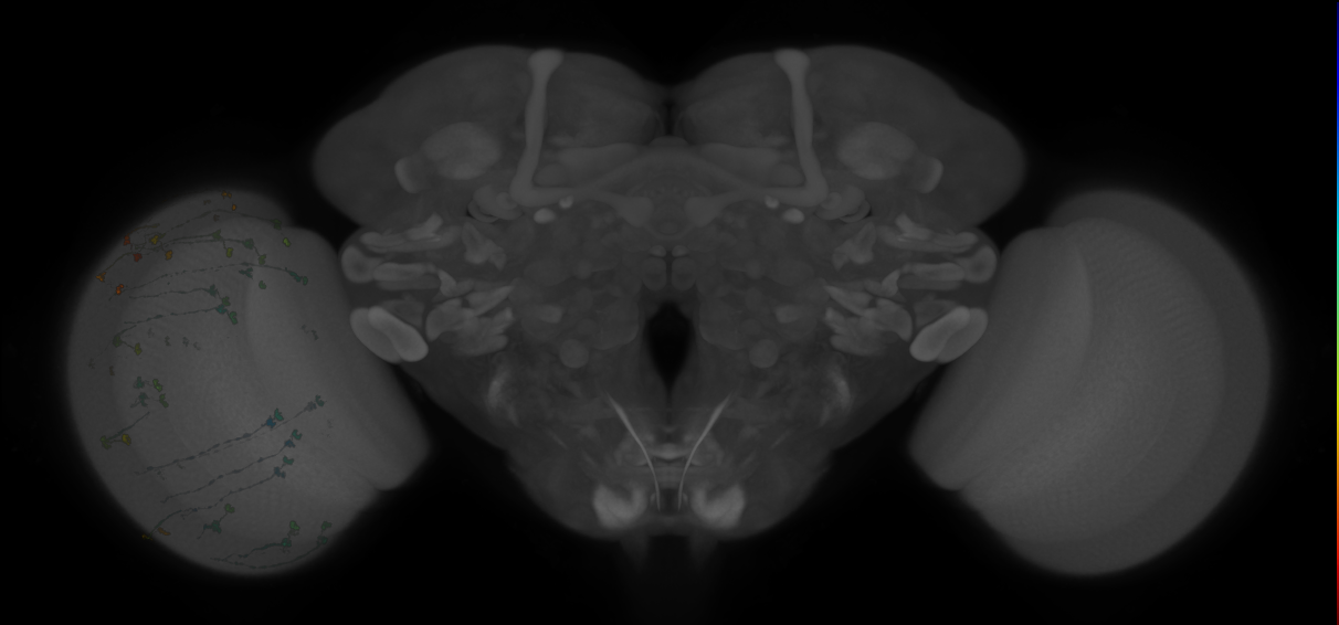
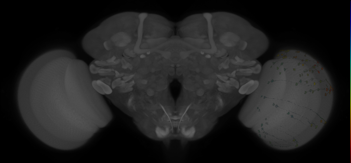
Split-GAL4 lines from Longden et al., 2023
ID: SplitLongden2023
Split-GAL4 lines from Longden et al., 2023
Open in VFB 3D Browser →Imaging Technique
- confocal microscopy
Downloads
Image files aligned to JRC_SS03696_Brain_20150821_20_F6_20x:
Image files aligned to JRC_SS03696_Brain_20150821_20_F5_20x:
References
161 - Split-GAL4 lines from Mais et al., 2021 [SplitMais2021]
Split-GAL4 lines from Mais et al., 2021
ID: SplitMais2021
Split-GAL4 lines from Mais et al., 2021
Open in VFB 3D Browser →References
162 - Split-GAL4 lines from Masek et al., 2015 [SplitMasek2015]
Split-GAL4 lines from Masek et al., 2015
ID: SplitMasek2015
Split-GAL4 lines from Masek et al., 2015
Open in VFB 3D Browser →Imaging Technique
- confocal microscopy
Downloads
Image files aligned to JRC_MB502B_Brain_20121210_33_D5_20x:
Image files aligned to JRC_MB502B_Brain_20121210_33_D4_63x:
Image files aligned to JRC_MB502B_Brain_20121210_33_D3_63x:
References
163 - Split-GAL4 lines from Meissner et al., 2018 [SplitMeissner2018]
Split-GAL4 lines from Meissner et al., 2018
ID: SplitMeissner2018
Split-GAL4 lines from Meissner et al., 2018
Open in VFB 3D Browser →References
164 - Split-GAL4 lines from Meissner et al., 2024 [SplitMeissner2024]
Split-GAL4 lines from Meissner et al., 2024
ID: SplitMeissner2024
Split-GAL4 lines from Meissner et al., 2024
Open in VFB 3D Browser →Imaging Technique
- confocal microscopy
Downloads
Image files aligned to JRC_SS55201_L3CNS_B:
Image files aligned to JRC_SS54251_L3CNS_B:
Image files aligned to JRC_SS53093_L3CNS_A:
Image files aligned to JRC_SS38412_L3CNS_A:
Image files aligned to JRC_SS38410_L3CNS_A:
Image files aligned to JRC_SS38049_L3CNS_A:
Image files aligned to JRC_SS42965_Brain_20171122_33_D5_20x:
Image files aligned to JRC_SS44402_Brain_20171108_31_B6_20x:
Image files aligned to JRC_SS43943_Brain_20171115_31_B4_63x:
Image files aligned to JRC_SS35366_VNC_20170531_33_A3_20x:
References
165 - Split-GAL4 lines from Minegishi et al., 2023 [SplitMinegishi2023]
Split-GAL4 lines from Minegishi et al., 2023
ID: SplitMinegishi2023
Split-GAL4 lines from Minegishi et al., 2023
Open in VFB 3D Browser →Imaging Technique
- confocal microscopy
Downloads
Image files aligned to JRC_SS25451_Brain_20171004_31_G2_20x:
Image files aligned to JRC_SS31456_Brain_20170816_31_D1_20x:
Image files aligned to JRC_SS25470_Brain_20170808_34_A3_20x:
Image files aligned to JRC_SS31456_VNC_20170816_31_D2_63x:
Image files aligned to JRC_SS25451_VNC_20171004_31_G3_20x:
Image files aligned to JRC_SS31480_VNC_20170816_32_C4_20x:
Image files aligned to JRC_SS25470_VNC_20170808_34_A2_20x:
Image files aligned to JRC_SS28596_VNC_20170726_32_A4_20x:
Image files aligned to JRC_SS28596_VNC_20170726_32_A2_20x:
Image files aligned to JRC_SS28596_VNC_20170726_32_A1_20x:
References
166 - Split-GAL4 lines from Montague et al., 2019 [SplitMontague2019]
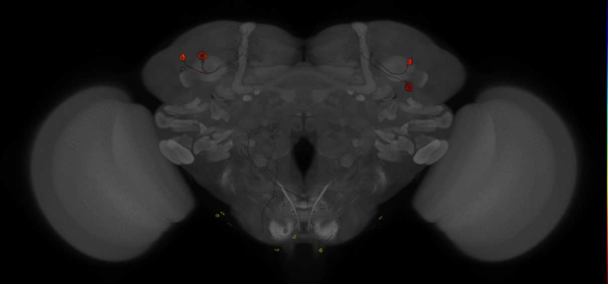
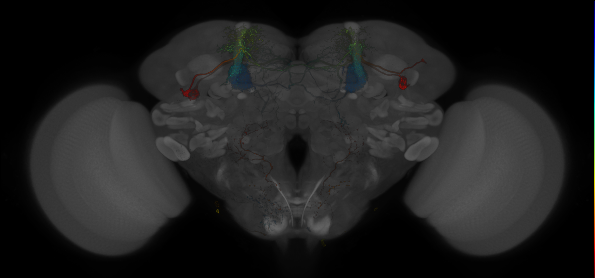
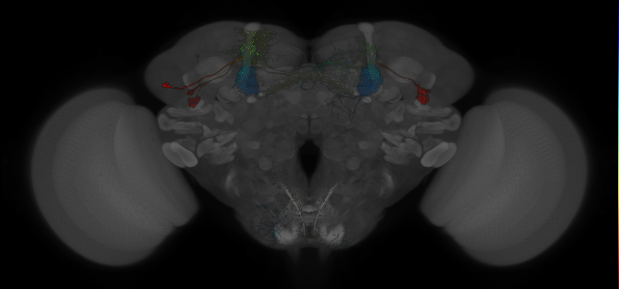
Split-GAL4 lines from Montague et al., 2019
ID: SplitMontague2019
Split-GAL4 lines from Montague et al., 2019
Open in VFB 3D Browser →Imaging Technique
- confocal microscopy
Downloads
Image files aligned to JRC_MB040B_Brain_20121212_31_D3_20x:
Image files aligned to JRC_MB439B_Brain_20121210_33_F3_20x:
Image files aligned to JRC_MB439B_Brain_20121108_19_H2_63x:
Image files aligned to JRC_MB439B_Brain_20121108_19_G2_63x:
Image files aligned to JRC_MB439B_Brain_20121108_19_G1_63x:
Image files aligned to JRC_MB439B_Brain_20121108_19_H1_20x:
Image files aligned to JRC_MB439B_Brain_20121108_19_G4_20x:
Image files aligned to JRC_MB040B_Brain_20121212_31_D2_63x:
Image files aligned to JRC_MB040B_Brain_20121212_31_D1_63x:
Image files aligned to JRC_MB439B_VNC_20121210_33_F1_63x:
References
167 - Split-GAL4 lines from Morimoto et al., 2020 [SplitMorimoto2020]
Split-GAL4 lines from Morimoto et al., 2020
ID: SplitMorimoto2020
Split-GAL4 lines from Morimoto et al., 2020
Open in VFB 3D Browser →References
168 - Split-GAL4 lines from Nallasivan et al 2025 [SplitNallasivan2025]


Split-GAL4 lines from Nallasivan et al 2025
ID: SplitNallasivan2025
Split-GAL4 lines from Nallasivan et al 2025
Open in VFB 3D Browser →Imaging Technique
- confocal microscopy
Downloads
Image files aligned to SPR8-Dsx SPRINs_VNC:
Image files aligned to SPR8-Fru11_SPRINs_VNC:
Image files aligned to SPR8-FD6 SPRINs_VNC:
Image files aligned to Fru11-FD6 SPRINs_VNC:
Image files aligned to Fru11-FD6 SPRINs_brain:
Image files aligned to SPR8-Fru11_SPRINs_brain:
References
169 - Split-GAL4 lines from Namiki et al., 2022 [SplitNamiki2022]
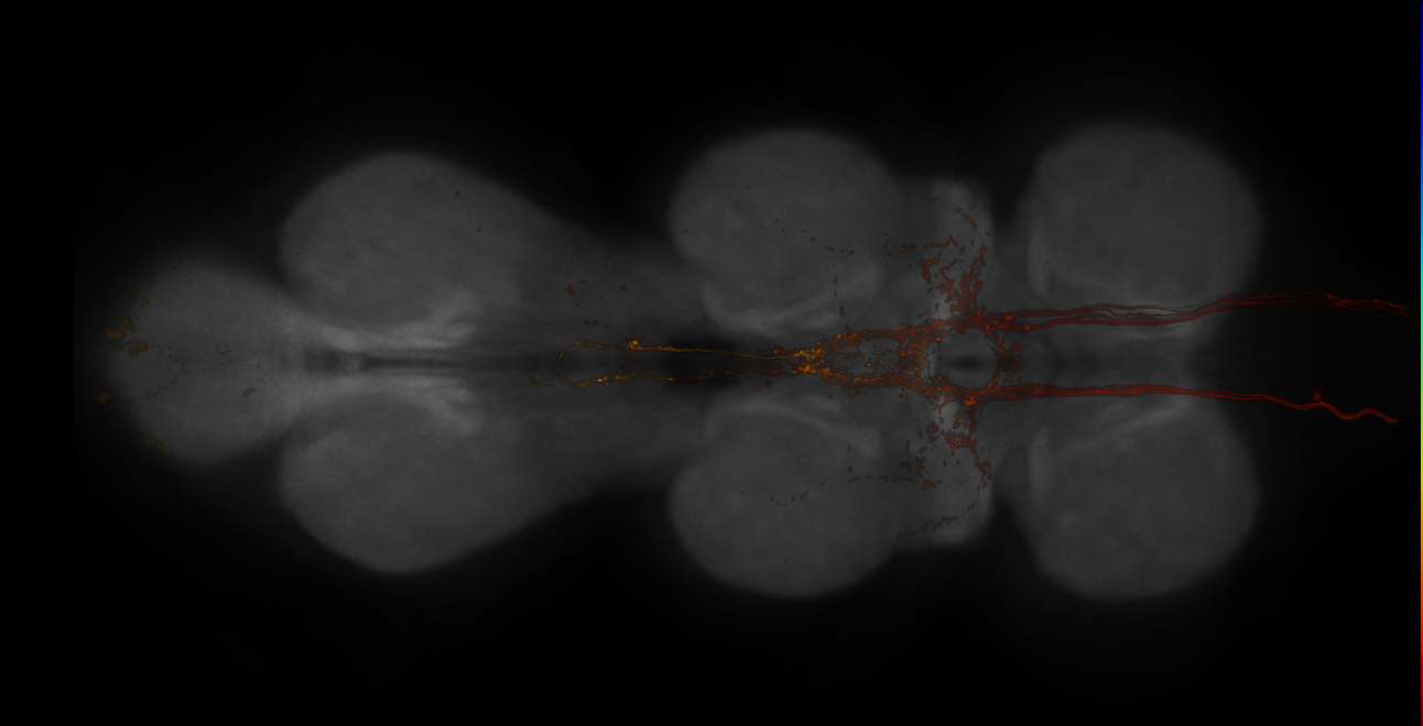
Split-GAL4 lines from Namiki et al., 2022
ID: SplitNamiki2022
Split-GAL4 lines labelling descending neurons for flight control from Namiki et al., 2022
Open in VFB 3D Browser →Imaging Technique
- confocal microscopy
Downloads
Image files aligned to JRC_SS01562_VNC_20140722_34_E1_63x:
Image files aligned to JRC_SS01563_VNC_20140819_33_D1_20x:
References
170 - Split-GAL4 lines from Nern et al., 2025 [SplitNern2025]
Split-GAL4 lines from Nern et al., 2025
ID: SplitNern2025
Split-GAL4 lines labelling optic lobe neurons from Nern et al., 2025
Open in VFB 3D Browser →References
171 - Split-GAL4 lines from Nojima et al., 2021 [Nojima2021]


Split-GAL4 lines from Nojima et al., 2021
ID: Nojima2021
Split-GAL4 lines labelling doublesex-expressing single neuronal clusters or neurons in the brain of both males and females from Nojima et al, 2021
Open in VFB 3D Browser →Imaging Technique
- confocal microscopy
Downloads
Image files aligned to Male_SN:
Image files aligned to Female_pMN2B:
Image files aligned to Male_pLN:
Image files aligned to Male_pC2m:
Image files aligned to Female_pCd-2:
Image files aligned to Male_pCd-2:
Image files aligned to Female_pCd-1:
Image files aligned to Female_pC2l:
Image files aligned to Female_pC1:
Image files aligned to Female_pMN2V:
References
172 - Split-GAL4 lines from Reyn et al., 2017 [SplitReyn2017]
Split-GAL4 lines from Reyn et al., 2017
ID: SplitReyn2017
Split-GAL4 lines from Reyn et al., 2017
Open in VFB 3D Browser →References
173 - Split-GAL4 lines from Ribeiro et al., 2018 [SplitRibeiro2018]
Split-GAL4 lines from Ribeiro et al., 2018
ID: SplitRibeiro2018
Split-GAL4 lines from Ribeiro et al., 2018
Open in VFB 3D Browser →References
174 - Split-GAL4 lines from Rubin et al., 2024 [SplitRubin2024]
Split-GAL4 lines from Rubin et al., 2024
ID: SplitRubin2024
Split-GAL4 lines from Rubin et al., 2024
Open in VFB 3D Browser →References
175 - Split-GAL4 lines from Schlichting et al., 2019 [SplitSchlichting2019]
Split-GAL4 lines from Schlichting et al., 2019
ID: SplitSchlichting2019
Split-GAL4 lines from Schlichting et al., 2019
Open in VFB 3D Browser →Imaging Technique
- confocal microscopy
Downloads
Image files aligned to JRC_SS00849_Brain_20170606_31_A5_20x:
Image files aligned to JRC_SS00849_Brain_20170606_31_A4_20x:
References
176 - Split-GAL4 lines from Schretter et al., 2020 [SplitSchretter2020]
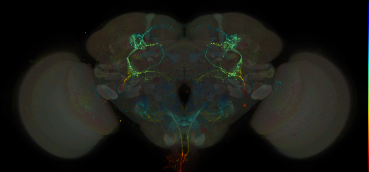
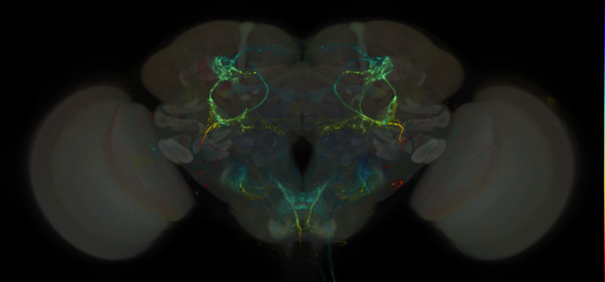
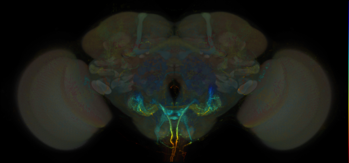
Split-GAL4 lines from Schretter et al., 2020
ID: SplitSchretter2020
Split-GAL4 lines from Schretter et al 2020
Open in VFB 3D Browser →Imaging Technique
- confocal microscopy
Downloads
Image files aligned to JRC_SS32237_Brain_20170223_33_F7_20x:
Image files aligned to JRC_SS32237_Brain_20170223_33_F3_20x:
Image files aligned to JRC_SS32237_Brain_20170223_33_F2_20x:
Image files aligned to JRC_SS32237_Brain_20170223_33_A6_20x:
Image files aligned to JRC_SS32237_Brain_20160708_27_H5_20x:
Image files aligned to SS32237:
Image files aligned to SS47478:
Image files aligned to SS39313:
Image files aligned to SS56964:
Image files aligned to SS36551:
References
177 - Split-GAL4 lines from Schretter et al., 2024 [SplitSchretter2024]
Split-GAL4 lines from Schretter et al., 2024
ID: SplitSchretter2024
Split-GAL4 lines labelling neurons which regulate visual processing from Schretter et al., 2024
Open in VFB 3D Browser →References
178 - Split-GAL4 lines from Shao et al., 2017 [SplitShao2017]
Split-GAL4 lines from Shao et al., 2017
ID: SplitShao2017
Split-GAL4 lines from Shao et al., 2017
Open in VFB 3D Browser →References
179 - Split-GAL4 lines from Shuai et al., 2023 [SplitShuai2023]
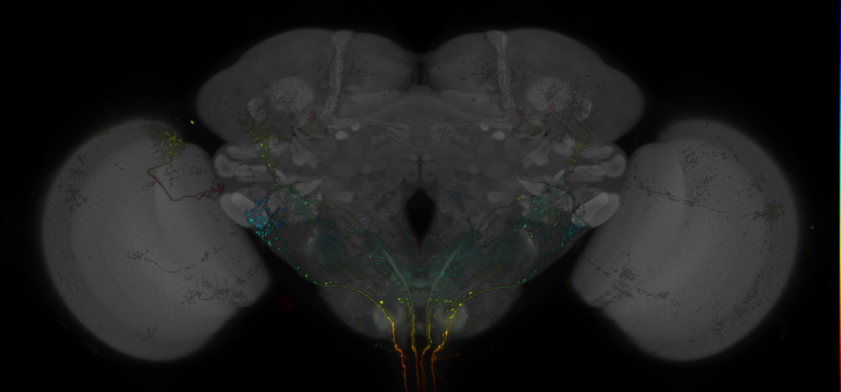
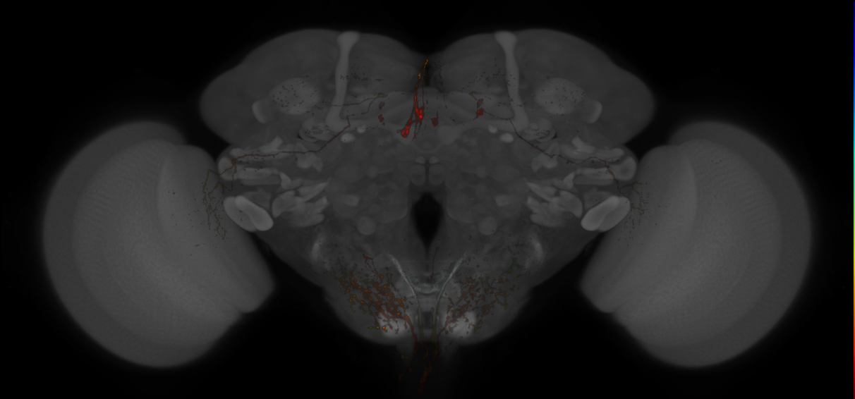
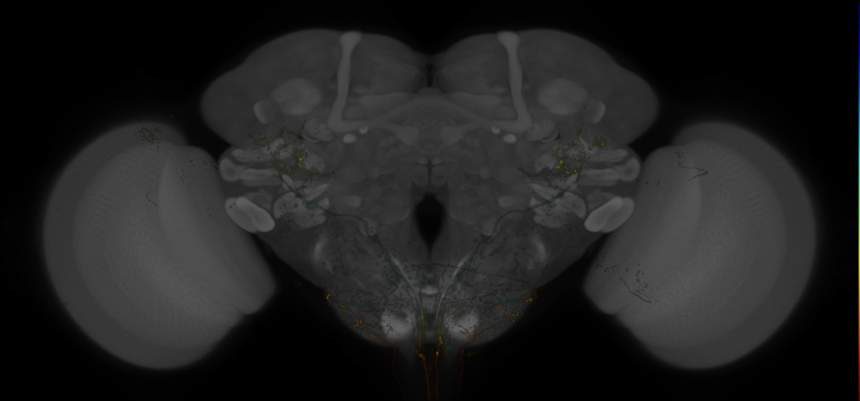
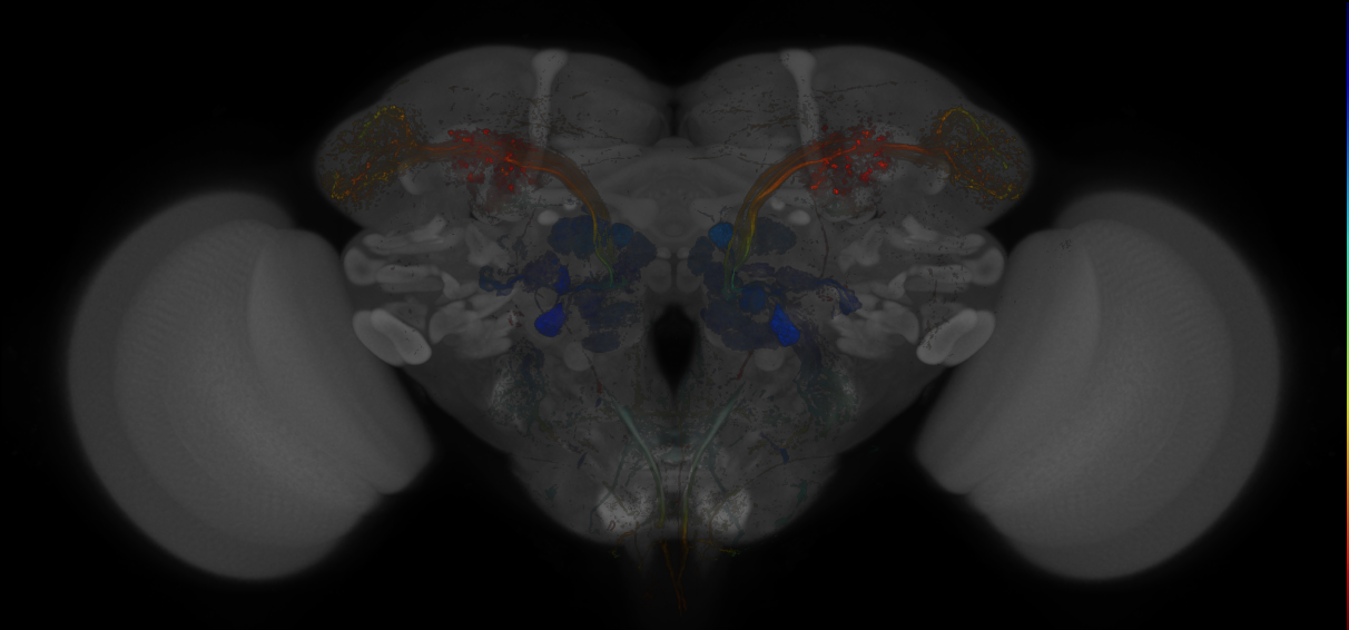
Split-GAL4 lines from Shuai et al., 2023
ID: SplitShuai2023
Split-GAL4 lines from Shuai et al., 2023
Open in VFB 3D Browser →Imaging Technique
- confocal microscopy
Downloads
Image files aligned to JRC_SS35253_Brain_20170103_31_I4_20x:
Image files aligned to JRC_SS11861_Brain_20170105_31_H1_20x:
Image files aligned to JRC_SS35241_Brain_20170105_31_F1_20x:
Image files aligned to JRC_SS32211_Brain_20160726_32_B3_20x:
Image files aligned to JRC_SS01214_Brain_20140808_32_I1_20x:
Image files aligned to JRC_SS01851_Brain_20140902_20_E1_63x:
Image files aligned to JRC_SS00589_Brain_20130204_19_I1_63x:
Image files aligned to JRC_SS45091_Brain_20171117_20_D7_20x:
Image files aligned to JRC_SS01168_Brain_20140825_19_E3_20x:
Image files aligned to JRC_SS00459_Brain_20130204_19_B2_20x:
References
180 - Split-GAL4 lines from Sitaraman et al., 2015 [SplitSitaraman2015]
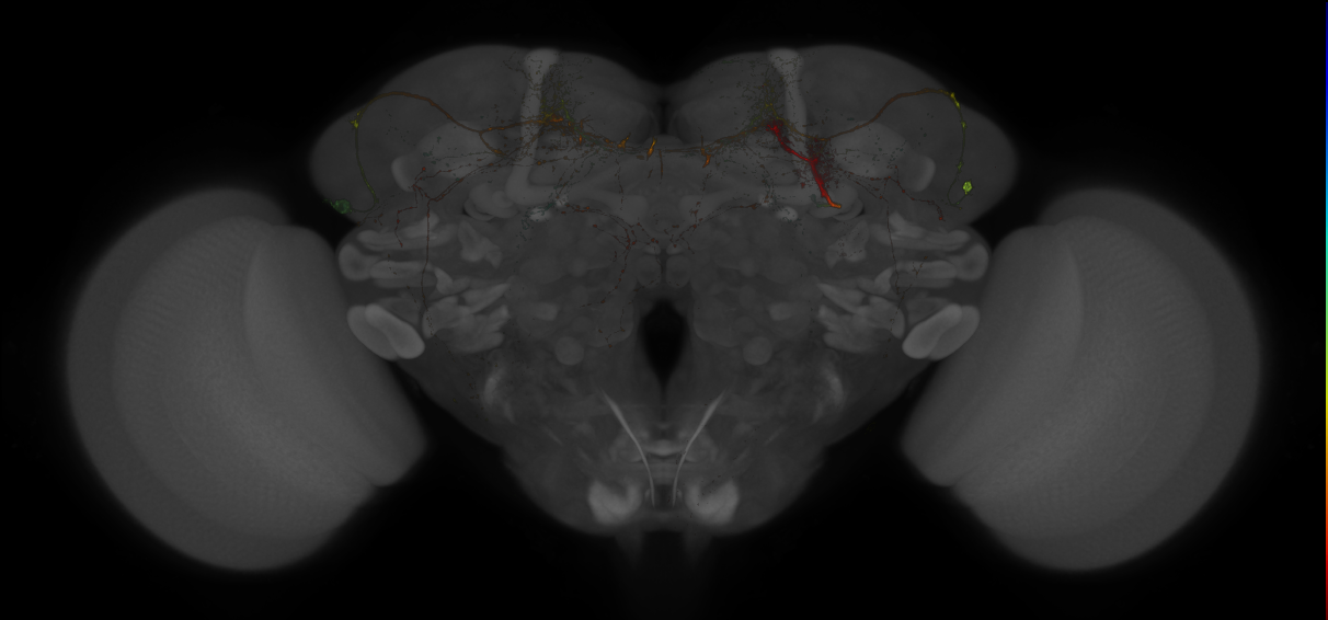
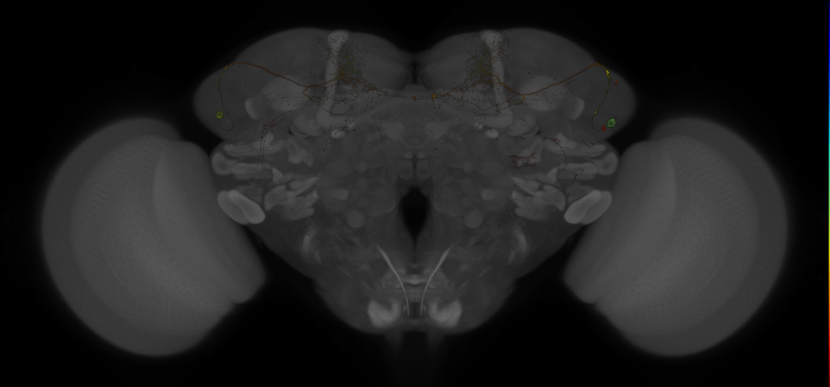
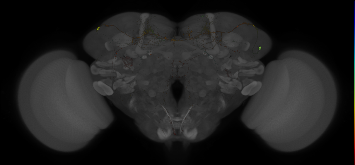
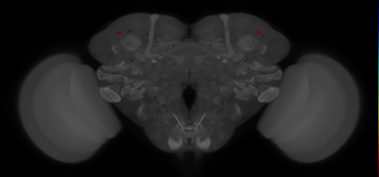
Split-GAL4 lines from Sitaraman et al., 2015
ID: SplitSitaraman2015
Split-GAL4 lines from Sitaraman et al., 2015
Open in VFB 3D Browser →Imaging Technique
- confocal microscopy
Downloads
Image files aligned to JRC_MB460B_Brain_20121208_32_A4_20x:
Image files aligned to JRC_MB460B_Brain_20121208_32_A3_20x:
Image files aligned to JRC_MB460B_Brain_20121208_32_A2_20x:
Image files aligned to JRC_MB052B_Brain_20121210_32_B2_20x:
Image files aligned to JRC_MB052B_Brain_20121210_32_A3_20x:
Image files aligned to JRC_MB460B_Brain_20121106_19_G3_63x:
Image files aligned to JRC_MB460B_Brain_20121106_19_H2_20x:
Image files aligned to JRC_MB460B_Brain_20121106_19_G4_20x:
Image files aligned to JRC_MB052B_Brain_20121210_32_B3_63x:
Image files aligned to JRC_MB052B_Brain_20121210_32_B1_63x:
References
181 - Split-GAL4 lines from Sterne et al., 2021 [SplitSterne2021]
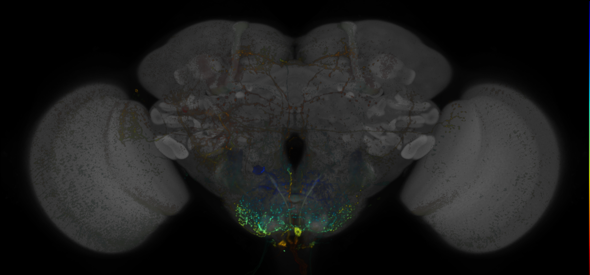
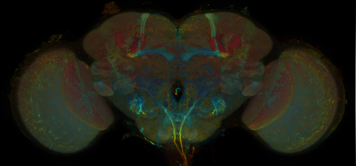
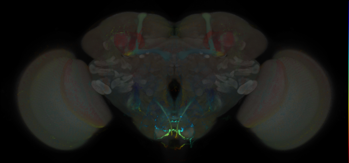
Split-GAL4 lines from Sterne et al., 2021
ID: SplitSterne2021
Split-GAL4 lines labelling adult subesophageal zone neurons from Sterne et al., 2021
Open in VFB 3D Browser →Imaging Technique
- confocal microscopy
Downloads
Image files aligned to JRC_SS42322_Brain_20171108_32_C5_20x:
Image files aligned to JRC_SS41389_Brain_20170927_31_B6_20x:
Image files aligned to JRC_SS49415_Brain_20170809_31_B4_20x:
Image files aligned to JRC_SS32361_Brain_20170621_32_A5_20x:
Image files aligned to JRC_SS29458_Brain_20170614_32_C3_20x:
Image files aligned to JRC_SS41410_Brain_20170906_34_J1_20x:
Image files aligned to JRC_SS28878_Brain_20171215_21_A1_20x:
Image files aligned to JRC_SS49415_Brain_20170707_25_C1_20x:
Image files aligned to JRC_SS35823_VNC_20170823_33_I2_20x:
Image files aligned to JRC_SS31341_VNC_20170628_31_H1_20x:
References
182 - Split-GAL4 lines from Sun et al., 2017 [SplitSun2017]
Split-GAL4 lines from Sun et al., 2017
ID: SplitSun2017
Split-GAL4 lines from Sun et al., 2017
Open in VFB 3D Browser →References
183 - Split-GAL4 lines from Takagi et al., 2017 [SplitTakagi2017]
Split-GAL4 lines from Takagi et al., 2017
ID: SplitTakagi2017
Split-GAL4 lines from Takagi et al., 2017
Open in VFB 3D Browser →References
184 - Split-GAL4 lines from Takemura et al., 2017 [SplitTakemura2017]
Split-GAL4 lines from Takemura et al., 2017
ID: SplitTakemura2017
Split-GAL4 lines from Takemura et al., 2017
Open in VFB 3D Browser →References
185 - Split-GAL4 lines from Tanaka et al., 2008 [SplitTanaka2008]
Split-GAL4 lines from Tanaka et al., 2008
ID: SplitTanaka2008
Split-GAL4 lines from Tanaka et al., 2008
Open in VFB 3D Browser →References
186 - Split-GAL4 lines from Triphan et al., 2016 [SplitTriphan2016]
Split-GAL4 lines from Triphan et al., 2016
ID: SplitTriphan2016
Split-GAL4 lines from Triphan et al., 2016
Open in VFB 3D Browser →References
187 - Split-GAL4 lines from Turner-Evans et al., 2017 [SplitDavis2017]
Split-GAL4 lines from Turner-Evans et al., 2017
ID: SplitDavis2017
Split-GAL4 lines from Davis et al 2017
Open in VFB 3D Browser →References
188 - Split-GAL4 lines from Turner-Evans et al., 2017 [SplitTurner_Evans2017]
Split-GAL4 lines from Turner-Evans et al., 2017
ID: SplitTurner_Evans2017
Split-GAL4 lines from Turner_Evans et al 2017
Open in VFB 3D Browser →References
189 - Split-GAL4 lines from Turner-Evans et al., 2020 [SplitTurner_Evans2020]
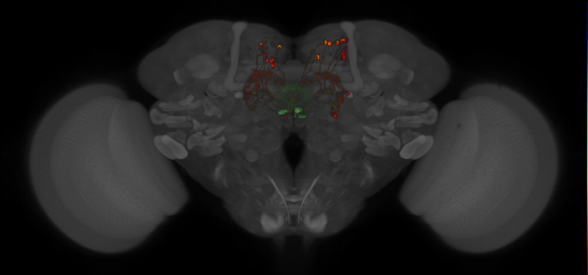
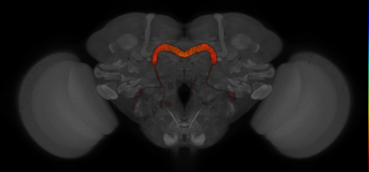
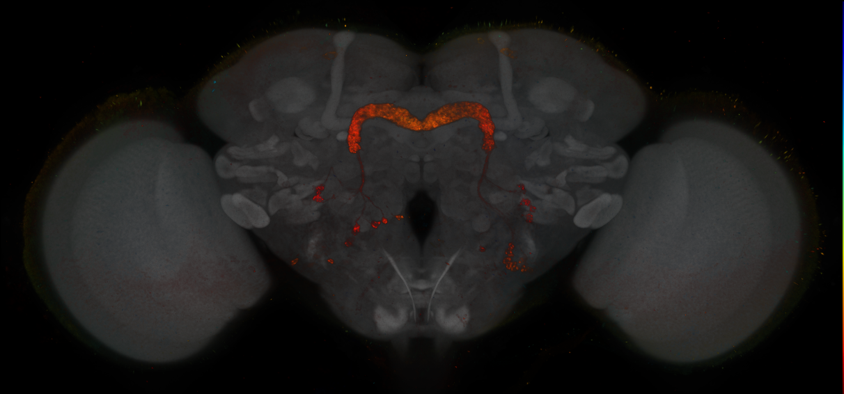

Split-GAL4 lines from Turner-Evans et al., 2020
ID: SplitTurner_Evans2020
Split-GAL4 lines from Turner_Evans et al 2020
Open in VFB 3D Browser →Imaging Technique
- confocal microscopy
Downloads
Image files aligned to SS62862:
Image files aligned to SS30295:
Image files aligned to SS02238:
Image files aligned to SS00238:
Image files aligned to SS02232:
Image files aligned to SS62862:
Image files aligned to SS02238:
Image files aligned to SS00238:
References
190 - Split-GAL4 lines from Tuthill et al., 2013 [SplitTuthill2013]
Split-GAL4 lines from Tuthill et al., 2013
ID: SplitTuthill2013
Split-GAL4 lines from Tuthill et al., 2013
Open in VFB 3D Browser →References
191 - Split-GAL4 lines from Vijayan et al., 2023 [SplitVijayan2023]
Split-GAL4 lines from Vijayan et al., 2023
ID: SplitVijayan2023
Split-GAL4 lines from Vijayan et al., 2023
Open in VFB 3D Browser →References
192 - Split-GAL4 lines from Vogt et al., 2014 [SplitVogt2014]
Split-GAL4 lines from Vogt et al., 2014
ID: SplitVogt2014
Split-GAL4 lines from Vogt et al., 2014
Open in VFB 3D Browser →References
193 - Split-GAL4 lines from Vogt et al., 2016 [SplitVogt2016]
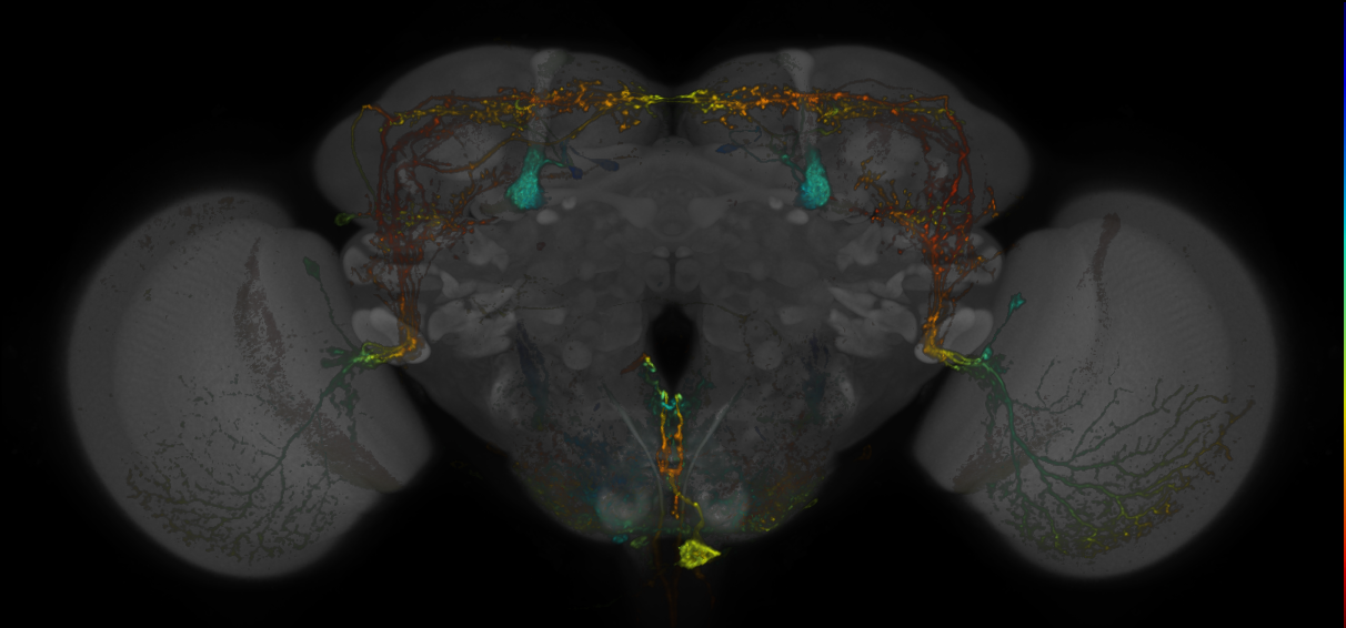
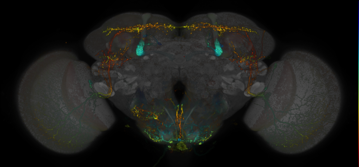
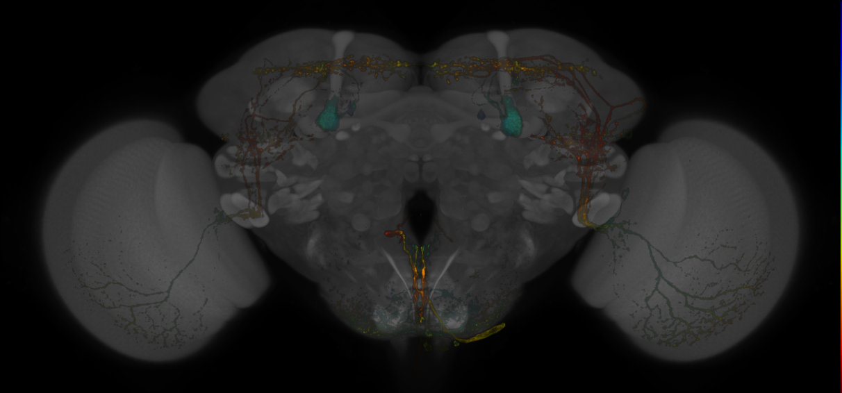
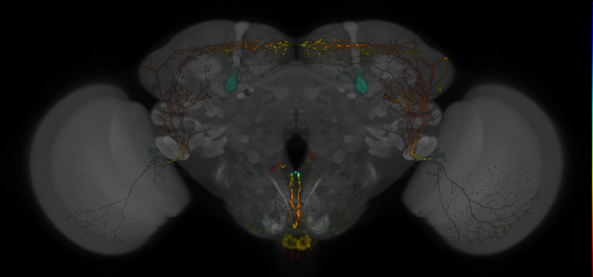
Split-GAL4 lines from Vogt et al., 2016
ID: SplitVogt2016
Split-GAL4 lines from Vogt et al., 2016
Open in VFB 3D Browser →Imaging Technique
- confocal microscopy
Downloads
Image files aligned to JRC_MB334C_Brain_20170829_34_F4_63x:
Image files aligned to JRC_MB334C_Brain_20170829_34_F6_20x:
Image files aligned to JRC_MB334C_Brain_20170829_34_F3_20x:
Image files aligned to JRC_MB334C_Brain_20170829_34_F2_20x:
Image files aligned to JRC_MB334C_Brain_20170829_34_E6_20x:
Image files aligned to JRC_MB334C_Brain_20170829_34_E5_20x:
Image files aligned to JRC_MB425B_Brain_20170829_34_D6_20x:
Image files aligned to JRC_MB425B_Brain_20170829_34_D5_20x:
Image files aligned to JRC_MB425B_Brain_20170829_34_D4_20x:
Image files aligned to JRC_MB425B_Brain_20170829_34_D3_20x:
References
194 - Split-GAL4 lines from Wang et al., 2020a [SplitWang2020a]
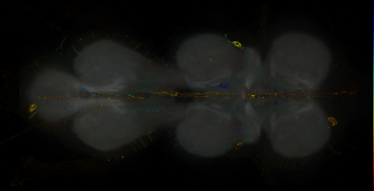
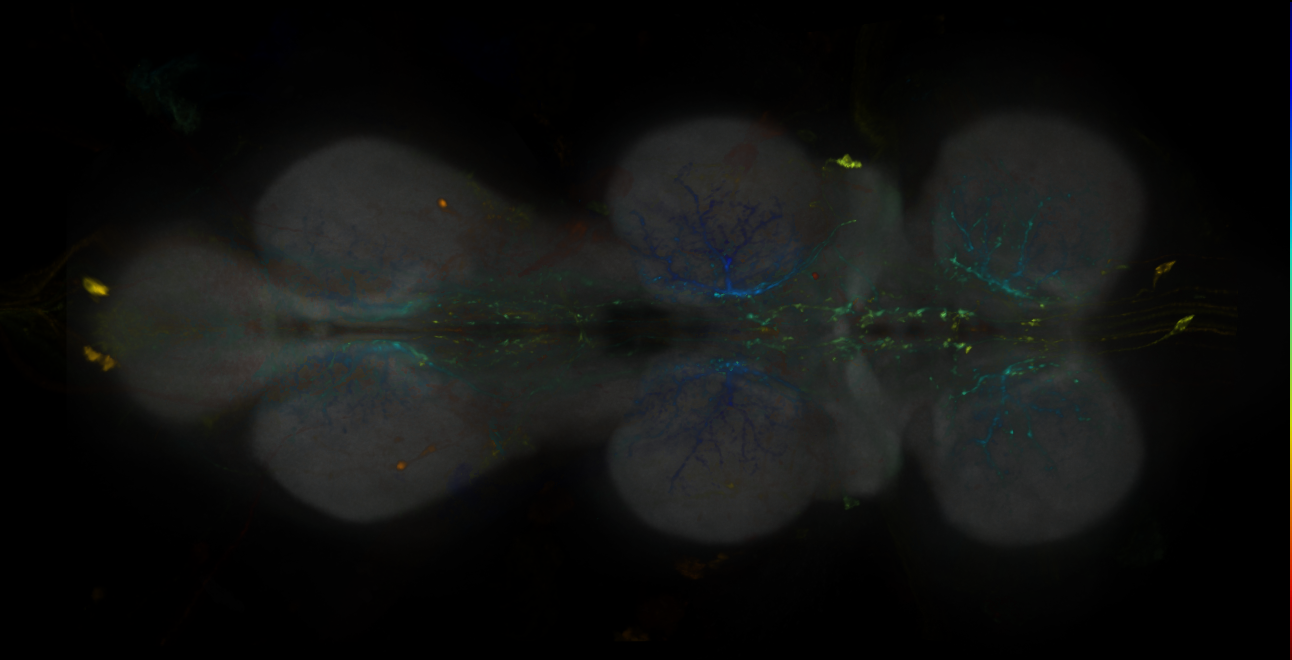
Split-GAL4 lines from Wang et al., 2020a
ID: SplitWang2020a
Split-GAL4 lines from Wang et al 2020
Open in VFB 3D Browser →Imaging Technique
- confocal microscopy
Downloads
Image files aligned to SS65426:
Image files aligned to SS51118:
Image files aligned to SS51118:
Image files aligned to SS50910:
Image files aligned to SS01491:
Image files aligned to SS59911:
Image files aligned to SS35666:
Image files aligned to SS35666:
Image files aligned to SS49443:
Image files aligned to SS51118:
References
195 - Split-GAL4 lines from Wang et al., 2020b [SplitWang2020b]
Split-GAL4 lines from Wang et al., 2020b
ID: SplitWang2020b
Split-GAL4 lines from Wang et al 2020b
Open in VFB 3D Browser →Imaging Technique
- confocal microscopy
Downloads
Image files aligned to SS60797:
Image files aligned to SS60797:
References
196 - Split-GAL4 lines from Wang et al., 2020c [SplitWang2020c]
Split-GAL4 lines from Wang et al., 2020c
ID: SplitWang2020c
Split-GAL4 lines labelling neurons that implement sexual receptivity decisions in Drosophila females from Wang et al., 2020c
Open in VFB 3D Browser →References
197 - Split-GAL4 lines from Wolff et al., 2025 [SplitWolff2025]
Split-GAL4 lines from Wolff et al., 2025
ID: SplitWolff2025
Split-GAL4 lines labelling central complex neurons from Wolff et al., 2025
Open in VFB 3D Browser →References
198 - Split-GAL4 lines from Xie et al., 2021 [SplitXie2021]
Split-GAL4 lines from Xie et al., 2021
ID: SplitXie2021
Split-GAL4 lines from Xie et al., 2021
Open in VFB 3D Browser →References
199 - Split-GAL4 lines from Yamagata et al., 2015 [SplitYamagata2015]
Split-GAL4 lines from Yamagata et al., 2015
ID: SplitYamagata2015
Split-GAL4 lines from Yamagata et al., 2015
Open in VFB 3D Browser →References
200 - Split-GAL4 lines from Yoo et al., 2023 [SplitYoo2023]
Split-GAL4 lines from Yoo et al., 2023
ID: SplitYoo2023
Split-GAL4 lines from Yoo et al., 2023
Open in VFB 3D Browser →References
201 - Split-GAL4 lines from Zhao et al., 2023 [SplitZhao2023]
Split-GAL4 lines from Zhao et al., 2023
ID: SplitZhao2023
Split-GAL4 lines from Zhao et al., 2023
Open in VFB 3D Browser →References
202 - Split-GAL4 lines from Zhao et al., 2025 [SplitZhao2025]
Split-GAL4 lines from Zhao et al., 2025
ID: SplitZhao2025
Split-GAL4 lines from Zhao et al., 2025
Open in VFB 3D Browser →References
203 - Split-GAL4 lines from Zung et al., 2025 [SplitZung2025]
Split-GAL4 lines from Zung et al., 2025
ID: SplitZung2025
Split-GAL4 lines labelling descending neurons from Zung et al., 2025
Open in VFB 3D Browser →References
204 - Splits targetting CX neurons, Wolff2018 [FlyLight2019Wolff2018]


Imaging Technique
- confocal microscopy
Downloads
Image files aligned to SS00098:
Image files aligned to SS52577:
Image files aligned to SS52267:
Image files aligned to SS50417:
Image files aligned to SS50417:
Image files aligned to SS54295:
Image files aligned to SS52577:
Image files aligned to SS52267:
Image files aligned to SS50417:
Image files aligned to SS50417:
References
205 - Splits targetting CX neurons, Wolff2018 [Wolff2018]
Imaging Technique
- confocal microscopy
Downloads
Image files aligned to SS00098:
Image files aligned to SS04912:
Image files aligned to SS02191:
Image files aligned to JRC_SS46528_Brain_20171128_31_I3_20x:
Image files aligned to SS50513:
Image files aligned to SS52631:
Image files aligned to SS47378:
Image files aligned to SS50517:
Image files aligned to SS47406:
Image files aligned to SS25983:
References
206 - Splits targetting the visual motion pathway, Strother2017 [FlyLight2019Strother2017]


Splits targetting the visual motion pathway, Strother2017
ID: FlyLight2019Strother2017
Imaging Technique
- confocal microscopy
Downloads
Image files aligned to SS31976:
Image files aligned to SS04528:
Image files aligned to SS01596:
Image files aligned to SS02555:
Image files aligned to SS02621:
Image files aligned to SS01594:
Image files aligned to SS01601:
Image files aligned to SS00737:
Image files aligned to SS00736:
Image files aligned to SS01070:
References
207 - Splits targetting the visual motion pathway, Strother2017 [Strother2017]
Splits targetting the visual motion pathway, Strother2017
ID: Strother2017
Imaging Technique
- confocal microscopy
Downloads
Image files aligned to SS02432:
Image files aligned to SS02430:
Image files aligned to SS00809:
Image files aligned to SS00300:
Image files aligned to SS00324:
Image files aligned to SS00313:
Image files aligned to SS00809:
Image files aligned to SS00300:
Image files aligned to SS02430:
Image files aligned to SS02432:
References
208 - Third order olfactory neurons involved in pheromone response - backfills (Kohl2013) [Kohl2013]


Third order olfactory neurons involved in pheromone response - backfills (Kohl2013)
ID: Kohl2013
Imaging Technique
- confocal microscopy
Downloads
Image files aligned to 120717-JK1029fill:
Image files aligned to 121122-2JK1029fill:
Image files aligned to 120817-JK56fill:
Image files aligned to 120812-2JK56fill:
Image files aligned to 120926-JK1029fill:
Image files aligned to 120717-JK1029fill:
Image files aligned to 121122-2JK1029fill:
Image files aligned to 120817-JK56fill:
Image files aligned to 120812-2JK56fill:
Image files aligned to 120926-JK1029fill:
References
209 - Truman Larval Flip-Out Collection [TrumanWood2018]


Truman Larval Flip-Out Collection
ID: TrumanWood2018
A selection of neurons from specimens created by Dr. Jim Truman at Janelia Research Campus using the multicolor flip out technique from the FlyLight GMR lines.
Open in VFB 3D Browser →Imaging Technique
- confocal microscopy
Downloads
Image files aligned to GMR 85G05 FLP 5-2:
Image files aligned to GMR 11H07 FLP 8-1:
Image files aligned to GMR 79G12 FLP 6-2:
Image files aligned to GMR 20F04 FLP 14-2:
Image files aligned to GMR 47D07 FLP 11-2:
Image files aligned to GMR 82F02 FLP 16-1:
Image files aligned to GMR 13B08 FLP 26-1:
Image files aligned to GMR 71E06 FLP 23-2:
Image files aligned to GMR 84B11 FLP 5-1:
Image files aligned to GMR 47H07 FLP 2-1:
References
210 - Truman Larval Flip-Out Collection [TrumanWood2018public]


Truman Larval Flip-Out Collection
ID: TrumanWood2018public
Images by James W. Truman and the Fly Light Project Team at HHMI Janelia Research Campus.
Open in VFB 3D Browser →Imaging Technique
- confocal microscopy
Downloads
Image files aligned to GMR 26H01 FLP 1-1:
Image files aligned to VT033614 FLP 4-2:
References
211 - Unveiling the sensory and interneuronal pathways of the neuroendocrine connectome in Drosophila (Hueckesfeld et al. 2020) [Hueckesfeld2020]


Unveiling the sensory and interneuronal pathways of the neuroendocrine connectome in Drosophila (Hueckesfeld et al. 2020)
ID: Hueckesfeld2020
L1 EM reconstructed neurons from Hueckesfeld et al. 2020
Open in VFB 3D Browser →Cross References
Imaging Technique
- transmission electron microscopy (TEM)
Downloads
Image files aligned to AN-L-Sens-B2-ACp-23 (L1EM:15997929):
Image files aligned to MN-R-Sens-B3-VM-09 (L1EM:15671848):
Image files aligned to _SOG IN (L1EM:14962387):
Image files aligned to MN-R-Sens-B1-ACpl-01 (L1EM:5448622):
Image files aligned to AN-L-Sens-B3-VM-12 (L1EM:5120097):
Image files aligned to neuron 19299842 (L1EM:19299841):
Image files aligned to putative MD III/IV_T1r (L1EM:360668):
Image files aligned to AF4: TOH left (L1EM:17641913):
Image files aligned to AN-L-Sens-B1-ACa-08 (L1EM:15769419):
Image files aligned to MB2ON-219 (L1EM:9455820):