OA-VUMa [FBbt_00110155]

OA-VUMa
ID: FBbt_00110155
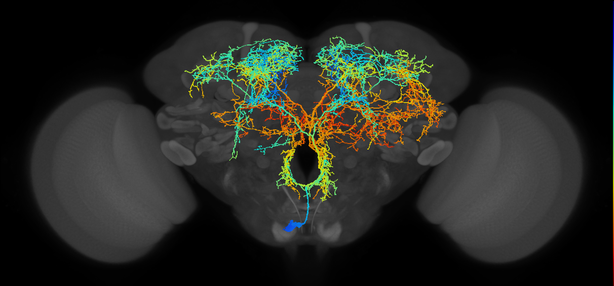
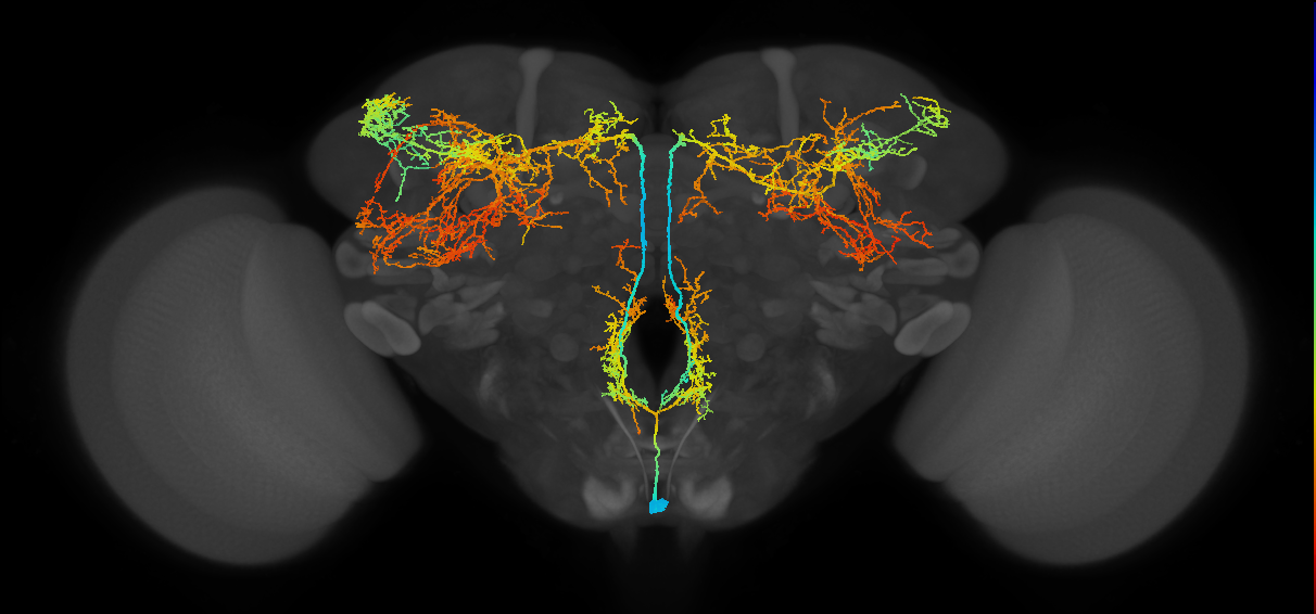
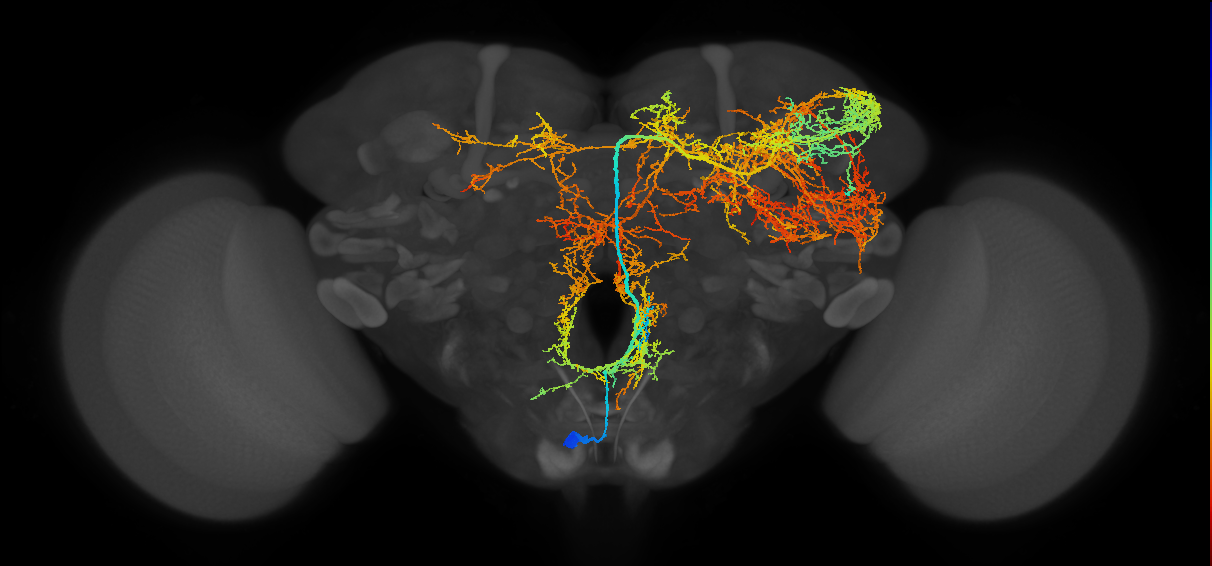
Octopaminergic neuron of the VUM cluster, whose cell body is located more anteriorly than those of OA-VUMd neurons, in either the mandibular or maxillary segments. Its primary neurite projects dorsally through the median tracts. It bifurcates in the area ventral to the esophagus foramen and form spiny dendritic arborizations in the posterior slope. Pairs of secondary processes emerge from these arbors and project to distinct brain regions, forming varicose nerve terminals (Busch et al., 2009, Busch and Tanimoto, 2010. (Busch et al., 2009, Busch and Tanimoto, 2010)
The neurotransmitter was assessed by immunostaining with an anti-octopamine (OA) antibody in cells labelled with tdc2-GAL4 (FBti0101786) and NP7088-GAL4 (FBti0037217) (Busch et al., 2009 or only using the tdc2-GAL4 (FBti0101786) (Busch and Tanimoto, 2010.
Open in VFB 3D Browser →Classification
Alternative Names
| Synonym | Scope | Reference |
|---|---|---|
| subesophageal ganglion ventral unpaired median neuron | broad synonym | Tanaka et al., 2008 |
| subesophageal ganglion VUM neuron | broad synonym | Tanaka et al., 2008 |
| ascending VUM neuron | broad synonym | Busch et al., 2009 |
| VUM neuron of the subesophageal ganglion | broad synonym | Tanaka et al., 2008 |
| OA-VUMa | exact synonym | Busch et al., 2009 |
| SOG VUM neuron | broad synonym | Tanaka et al., 2008 |
| ventral unpaired median octopaminergic anterior neuron | exact synonym |
Downloads
Image files aligned to OA-VUMa6(OA-VUMa7)_M (MaleCNS:10168):
Image files aligned to OA-VUMa3_M (MaleCNS:10555):
Image files aligned to OA-VUMa3_M (MaleCNS:10080):
Image files aligned to OA-VUMa6_R:
Image files aligned to OA-VUMa3:
Image files aligned to OA-VUMa3:
Image files aligned to BANC_626:720575941414866833:
Image files aligned to BANC_626:720575941492310759:
Image files aligned to OA-VUMa3_M (MaleCNS:10555):
Image files aligned to OA-VUMa6_R:
Graphs For
References
Feedback
Was this page helpful?
Glad to hear it! Please tell us how we can improve.
Sorry to hear that. Please tell us how we can improve.