OA-VL [FBbt_00110164]
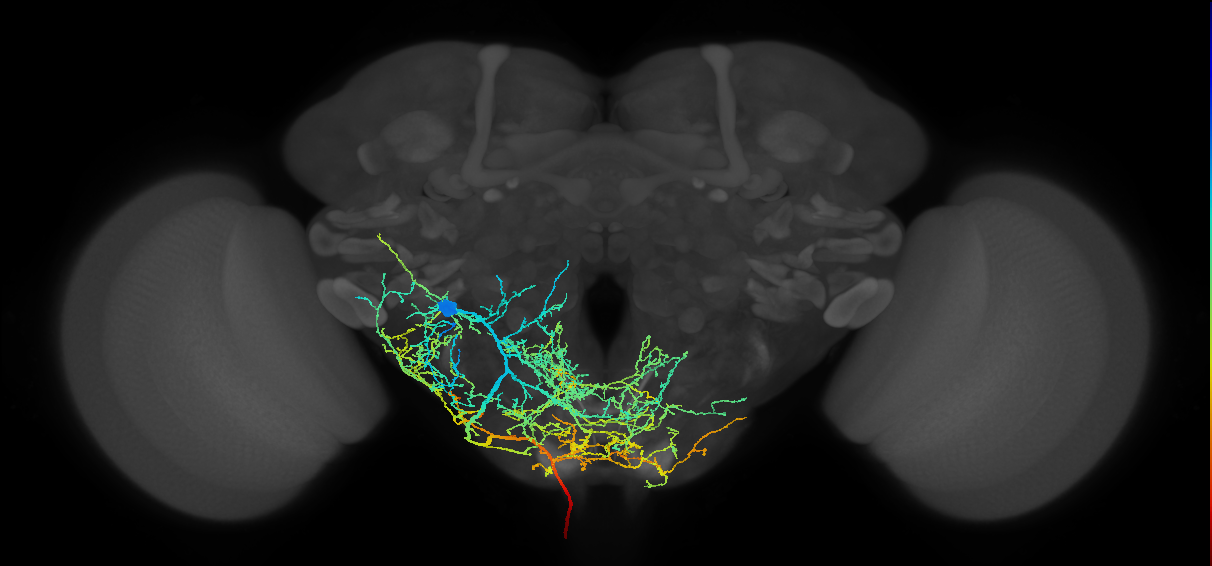
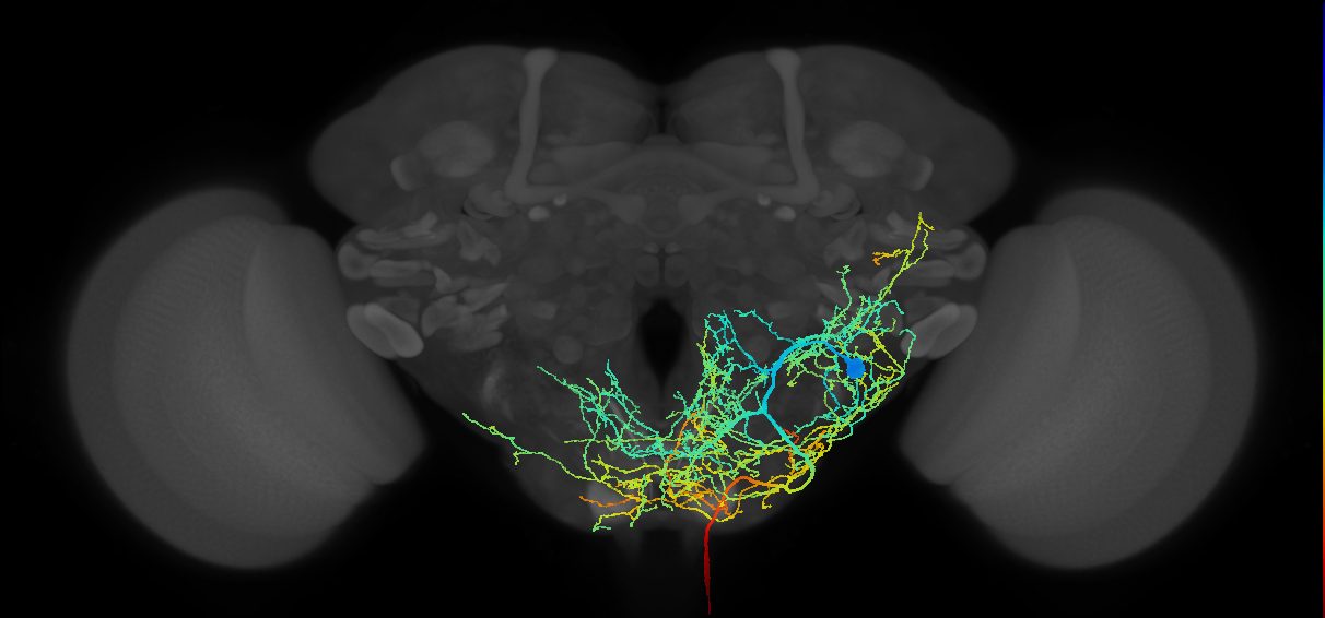


OA-VL
ID: FBbt_00110164
Octopaminergic neuron of the VL cluster, whose cell body is located between the antennal lobe and the ventrolateral protocerebrum of the adult brain. This neuron sends a process to the subesophageal zone where it crosses the midline. Bilateral ramifications arborize in the antennal lobes and the gnathal ganglion. On the ipsilateral side, ramifications extend to the inferior lateral and ventrolateral protocerebra. A descending axon diverges from the main neurite medial to the antennal nerve. Two different subtypes can be distinguished, which differ on the projection pattern of this descending neurite: OA-VL1 and OA-VL2. There are two neurons per cluster (Busch et al., 2009. (Busch et al., 2009)
The neurotransmitter was assessed by immunostaining with an anti-octopamine (OA) antibody in cells labelled with tdc2-GAL4 (FBti0101786) and NP7088-GAL4 (FBti0037217) (Busch et al., 2009. Both subtypes correspond to descending neurons of the outside anterior cluster (Sturner et al., 2025 - FlyBase:FBrf0262850).
Open in VFB 3D Browser →Classification
Relationships
- bearer of: bilateral
- has synaptic IO in region: adult protocerebrum, adult subesophageal zone
Alternative Names
| Synonym | Scope | Reference |
|---|---|---|
| OA-VL | exact synonym | Busch et al., 2009 |
| ventrolateral protocerebrum octopaminergic neuron | exact synonym | Busch et al., 2009 |
Downloads
Image files aligned to DNd03_L (MaleCNS:11486):
Image files aligned to DNd03_R (MaleCNS:11693):
Image files aligned to DNd03:
Image files aligned to DNd03:
Image files aligned to DNd02:
Image files aligned to DNd03_L (MaleCNS:11486):
Image files aligned to DNd03_R (MaleCNS:11693):
Image files aligned to TBD (MANC:10526):
Image files aligned to TBD (MANC:10491):
Image files aligned to TBD (MANC:10410):
Graphs For
References
Feedback
Was this page helpful?
Glad to hear it! Please tell us how we can improve.
Sorry to hear that. Please tell us how we can improve.