OA-AL2 [FBbt_00110139]


OA-AL2
ID: FBbt_00110139
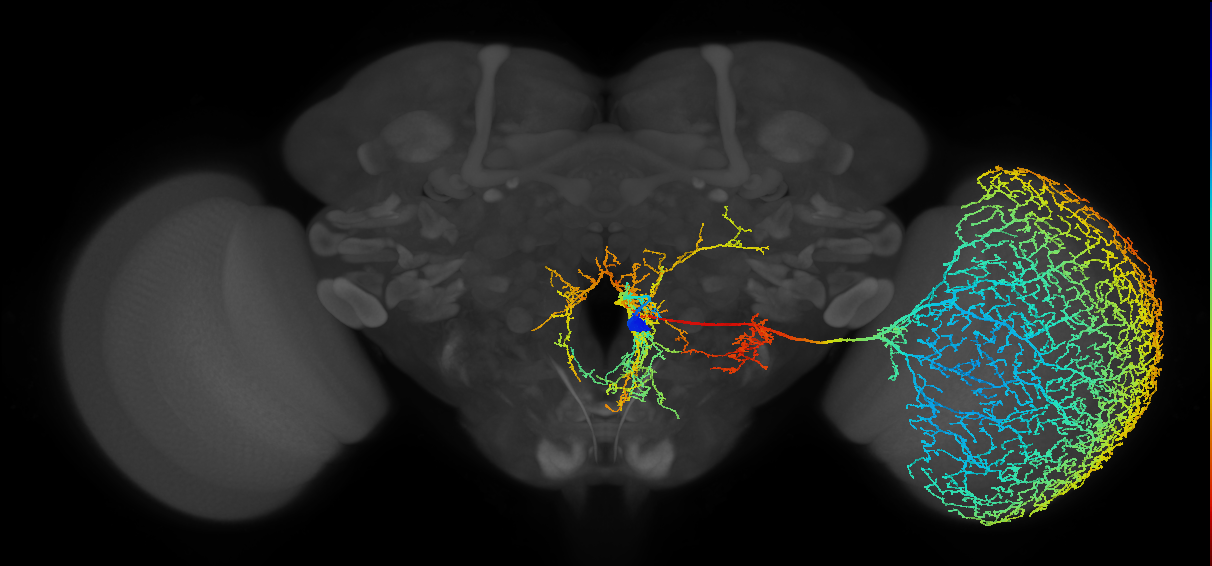
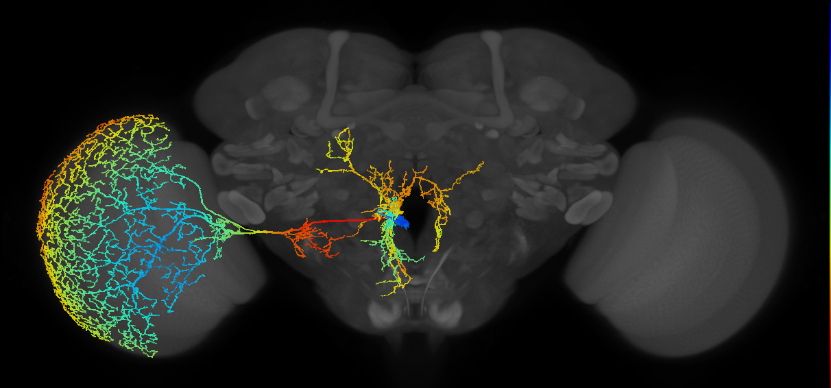
Octopaminergic neuron whose cell body is located in a cluster in the ventromedial border of the antennal lobe. One neurite runs along the esophagus foramen to the posterior slope where it branches, forming spiny dendritic arborizations. It has axonal projections that innervate the optic lobe. There are seven cells in this cluster that belong to two main subtypes, OA-AL2i and AL2b (Busch et al., 2009. (Busch et al., 2009)
The neurotransmitter was assessed by immunostaining with an anti-octopamine (OA) antibody in cells labelled with tdc2-GAL4 (FBti0101786) and NP7088-GAL4 (FBti0037217). Post- versus pre-synaptic innervation was determined using the postsynaptic marker Rdl-HA or the presynaptic marker Synaptotagmin-HA (Busch et al., 2009.
Open in VFB 3D Browser →Classification
Relationships
- has soma location: cell body rind of adult antennal lobe
- receives synaptic input in region: posterior slope
- sends synaptic output to region: adult optic lobe
Alternative Names
| Synonym | Scope | Reference |
|---|---|---|
| OA-AL2 | exact synonym | Busch et al., 2009 |
| octopaminergic neuron of the antennal lobe | exact synonym |
Downloads
Image files aligned to OA-AL2i3_R (MaleCNS:10825):
Image files aligned to OA-AL2i3_L (MaleCNS:10658):
Image files aligned to OA-AL2i3:
Image files aligned to OA-AL2i3:
Image files aligned to OA-AL2i3:
Image files aligned to BANC_626:720575941436697632:
Image files aligned to BANC_626:720575941593346155:
Image files aligned to BANC_626:720575941632601324:
Image files aligned to OA-AL2i3_R (JRC_OpticLobe:10825):
Image files aligned to OA-AL2i3_R (MaleCNS:10825):
Graphs For
References
Feedback
Was this page helpful?
Glad to hear it! Please tell us how we can improve.
Sorry to hear that. Please tell us how we can improve.