Dm10 FBbt_00110135
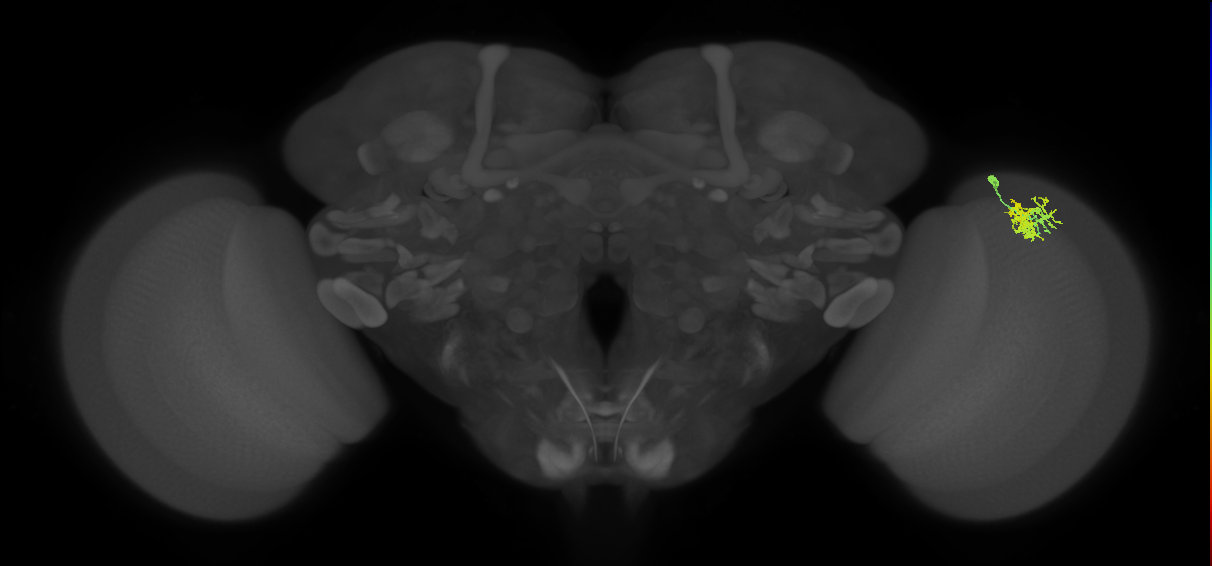
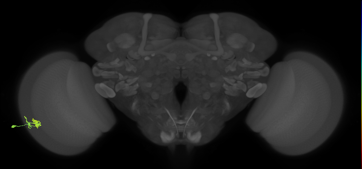


Dm10
ID: FBbt_00110135
Distal medullary wide-field amacrine neuron whose cell body is located in the cell body rind of the medulla. It has a distinct tri-stratified layer pattern, branching extensively in medulla layers M1, M3B and M5. Its arbor resembles an ellipse, with the long axis aligned with the dorsal-ventral axis, and covering less than 10 columns. The processes in M1 and M3B to M5 are located in an intracolumnar position. There are around 300 Dm10 neurons per hemisphere. They are GABAergic (Davis et al., 2020. (Takemura et al., 2013, Davis et al., 2020, Nern et al., 2015, Morante and Desplan, 2008)
Pre-synaptic terminals were assessed by labelling with a UAS-Synaptotagmin reporter (P{UAS-HA-syt} - FBtp0015803). The identification of dendrites and axons was done using a UAS-Dscam (P{UAS-Dscam.exon17.1.GFP} - FBtp0021447) or a UAS-tau (P{SUAS-tau-lacZ.O} - FBtp0010033) reporter, respectively (Morante and Desplan, 2008.
Open in VFB 3D Browser →Classification
- Notch OFF hemilineage secondary neuron
- adult GABAergic neuron
- distal medullary amacrine neuron
- optic lobe wide-field columnar neuron
Relationships
- develops from: immature distal medullary amacrine neuron Dm10
- receives synaptic input in region: medulla layer M1, medulla layer M5, medulla sublayer M3B
- sends synaptic output to region: medulla layer M1, medulla layer M5, medulla sublayer M3B
Alternative Names
| Synonym | Scope | Reference |
|---|---|---|
| Dm10 | related synonym | Bates et al., 2025, Berg et al., 2025, name_in_male-cns, Nern et al., 2025, Schlegel et al., 2024 |
| distal medullary amacrine neuron Dm1-5 | exact synonym | Morante and Desplan, 2008 |
| Dm1-5 | exact synonym | Morante and Desplan, 2008 |
| Dm10 | exact synonym | Özel et al., 2021 |
Downloads
Image files aligned to Dm10_R (MaleCNS:73391):
Image files aligned to Dm10_L (MaleCNS:131913):
Image files aligned to Dm10:
Image files aligned to Dm10:
Image files aligned to Dm10:
Image files aligned to Dm10:
Image files aligned to Dm10:
Image files aligned to Dm10:
Image files aligned to Dm10_R (JRC_OpticLobe:81016):
Image files aligned to Dm10_R (JRC_OpticLobe:50086):