Dm1 FBbt_00003768
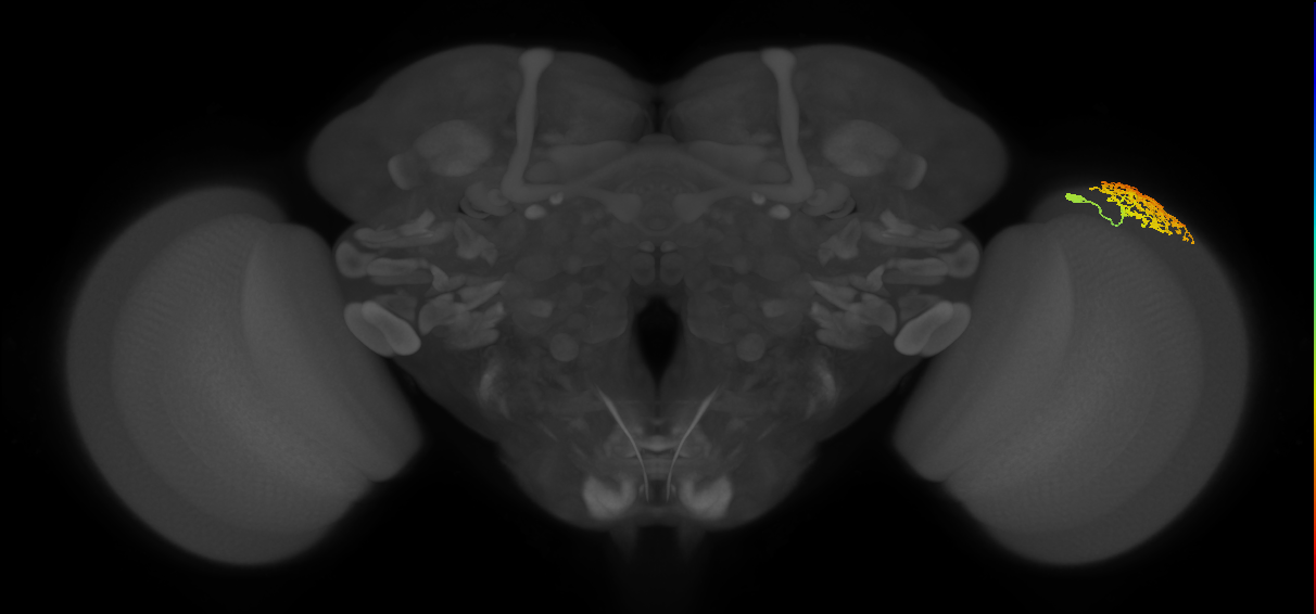
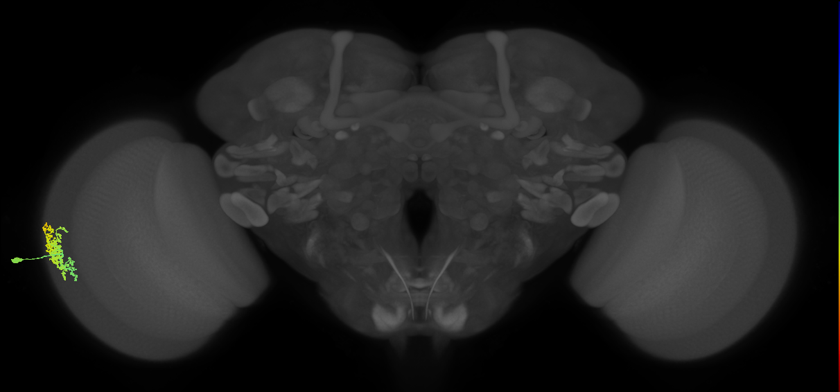
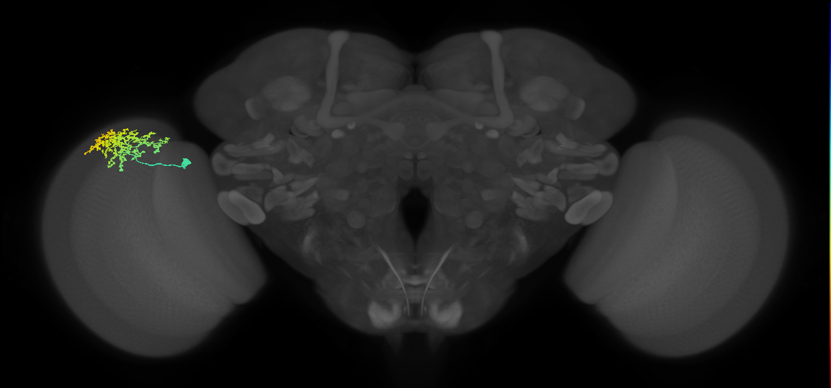

Dm1
ID: FBbt_00003768
Distal medullary wide-field amacrine neuron whose cell body is located in the anterior region of the cell body rind of the medulla. It branches extensively at the distal surface of the medulla forming a moderately broad arbor with each branch making a distinctive bouton-like terminal in the region between M1 and M2 (in the same sublayer as Dm18, and more proximal than Dm9 and Dm10 in M1), from which short, fine terminal branches project (Morante and Desplan, 2008; Fischbach and Dittrich, 1989. The arbor varies in shape between cells, covering around 20-30 columns, but with these areas overlapping. The size of the terminals of Dm1 is smaller than those of Dm18. There are around 40 Dm1 neurons per hemisphere. They are glutamatergic (Davis et al., 2020. (Davis et al., 2020, Fischbach and Dittrich, 1989, Morante and Desplan, 2008, Nern et al., 2015)
Pre- versus postsynaptic innervation judged by scoring of terminal morphology from figures in Fischbach and Dittrich, 1989 as assessed by FlyBrain Neuron DB.
Open in VFB 3D Browser →Classification
Relationships
- develops from: immature distal medullary amacrine neuron Dm1
- receives synaptic input in region: medulla layer M1
- sends synaptic output to region: medulla layer M1
Alternative Names
| Synonym | Scope | Reference |
|---|---|---|
| Dm1 | related synonym | Berg et al., 2025, Bates et al., 2025, name_in_male-cns, Schlegel et al., 2024, Nern et al., 2025 |
| Dm1 | exact synonym | Özel et al., 2021 |
Cross References
- Dm1 on FlyBrain NeuronDB (10081)
Downloads
Image files aligned to Dm1_R (MaleCNS:31091):
Image files aligned to Dm1_L (MaleCNS:559915):
Image files aligned to Dm1_L (MaleCNS:338369):
Image files aligned to Dm1:
Image files aligned to Dm1:
Image files aligned to Dm1:
Image files aligned to Dm1:
Image files aligned to Dm1_R (JRC_OpticLobe:27538):
Image files aligned to Dm1_R (JRC_OpticLobe:20806):
Image files aligned to Dm1_R (MaleCNS:31091):